Structure and Topology of the Huntingtin 1-17 Membrane Anchor by a Combined Solution and Solid-State NMR Approach.
Michalek, M., Salnikov, E.S., Bechinger, B.(2013) Biophys J 105: 699-710
- PubMed: 23931318
- DOI: https://doi.org/10.1016/j.bpj.2013.06.030
- Primary Citation of Related Structures:
2LD0, 2LD2 - PubMed Abstract:
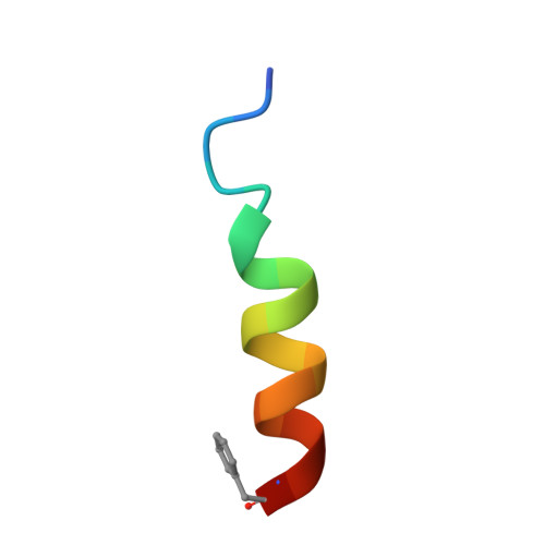
The very amino-terminal domain of the huntingtin protein is directly located upstream of the protein's polyglutamine tract, plays a decisive role in several important properties of this large protein and in the development of Huntington's disease. This huntingtin 1-17 domain is on the one hand known to markedly increase polyglutamine aggregation rates and on the other hand has been shown to be involved in cellular membrane interactions. Here, we determined the high-resolution structure of huntingtin 1-17 in dodecyl phosphocholine micelles and the topology of its helical domain in oriented phosphatidylcholine bilayers. Using two-dimensional solution NMR spectroscopy the low-energy conformations of the polypeptide were identified in the presence of dodecyl phosphocholine detergent micelles. In a next step a set of four solid-state NMR angular restraints was obtained from huntingtin 1-17 labeled with (15)N and (2)H at selected sites. Of the micellar ensemble of helical conformations only a limited set agrees in quantitative detail with the solid-state angular restraints of huntingtin 1-17 obtained in supported planar lipid bilayers. Thereby, the solid-state NMR data were used to further refine the domain structure in phospholipid bilayers. At the same time its membrane topology was determined and different motional regimes of this membrane-associated domain were explored. The pronounced structural transitions of huntingtin 1-17 upon membrane-association result in a α-helical conformation from K6 to F17, i.e., up to the very start of the polyglutamine tract. This amphipathic helix is aligned nearly parallel to the membrane surface (tilt angle ∼77°) and is characterized by a hydrophobic ridge on one side and an alternation of cationic and anionic residues that run along the hydrophilic face of the helix. This arrangement facilitates electrostatic interactions between huntingtin 1-17 domains and possibly with the proximal polyglutamine tract.
Organizational Affiliation:
Université de Strasbourg/CNRS, UMR, Institut de Chimie, France.