SARS coronavirus unique domain: three-domain molecular architecture in solution and RNA binding.
Johnson, M.A., Chatterjee, A., Neuman, B.W., Wuthrich, K.(2010) J Mol Biol 400: 724-742
- PubMed: 20493876
- DOI: https://doi.org/10.1016/j.jmb.2010.05.027
- Primary Citation of Related Structures:
2KAF, 2KQV, 2KQW - PubMed Abstract:
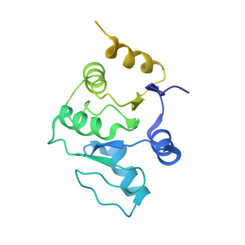
Nonstructural protein 3 of the severe acute respiratory syndrome (SARS) coronavirus includes a "SARS-unique domain" (SUD) consisting of three globular domains separated by short linker peptide segments. This work reports NMR structure determinations of the C-terminal domain (SUD-C) and a two-domain construct (SUD-MC) containing the middle domain (SUD-M) and the C-terminal domain, and NMR data on the conformational states of the N-terminal domain (SUD-N) and the SUD-NM two-domain construct. Both SUD-N and SUD-NM are monomeric and globular in solution; in SUD-NM, there is high mobility in the two-residue interdomain linking sequence, with no preferred relative orientation of the two domains. SUD-C adopts a frataxin like fold and has structural similarity to DNA-binding domains of DNA-modifying enzymes. The structures of both SUD-M (previously determined) and SUD-C (from the present study) are maintained in SUD-MC, where the two domains are flexibly linked. Gel-shift experiments showed that both SUD-C and SUD-MC bind to single-stranded RNA and recognize purine bases more strongly than pyrimidine bases, whereby SUD-MC binds to a more restricted set of purine-containing RNA sequences than SUD-M. NMR chemical shift perturbation experiments with observations of (15)N-labeled proteins further resulted in delineation of RNA binding sites (i.e., in SUD-M, a positively charged surface area with a pronounced cavity, and in SUD-C, several residues of an anti-parallel beta-sheet). Overall, the present data provide evidence for molecular mechanisms involving the concerted actions of SUD-M and SUD-C, which result in specific RNA binding that might be unique to the SUD and, thus, to the SARS coronavirus.
Organizational Affiliation:
Department of Molecular Biology, The Scripps Research Institute, La Jolla, CA 92037, USA.