Characterization of DLC1-SAM equilibrium unfolding at the amino acid residue level
Yang, S., Noble, C.G., Yang, D.(2009) Biochemistry 48: 4040-4049
- PubMed: 19317456
- DOI: https://doi.org/10.1021/bi9000936
- Primary Citation of Related Structures:
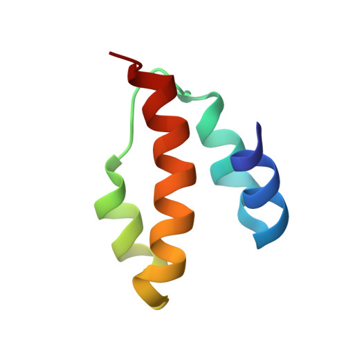
2KAP - PubMed Abstract:
Sterile alpha motif (SAM) domains are found in many different proteins and shown to play important roles in various biological processes. The N-terminal domain of deleted in liver cancer 1 (DLC1) protein is a SAM domain which exists in a monomeric form in aqueous solution and facilitates the distribution of EF1A1 to the membrane periphery and ruffles upon growth factor stimulation. Here, we report the structure of an N-terminal truncated DLC1 SAM domain (DLC1-SAM) and its urea-induced equilibrium unfolding investigated with various biophysical methods such as CD, fluorescence emission spectroscopy, and NMR. CD and tryptophan intrinsic fluorescence emission data imply that the unfolding of DLC1-SAM follows a simple two-state mechanism, yet the NMR data suggest the presence of at least one intermediate state. The intermediate cannot be detected by NMR, but it does not exist in large aggregates as shown by analytical ultracentrifugation experiments. Analysis of the free energy values for different residues shows that in the transition from the native state to non-native states the C-terminal helix is somewhat more stable than the other parts of the protein, whereas in the transition from the native and intermediate states to the denatured state, the stabilities of different residues are similar except for that of the region surrounding residues D37-F40 which has lower stability and is more readily denatured at high urea concentrations. Analysis of the midpoints of the transitions shows that the unfolding of the native state and formation of the denatured state are not cooperative and the unfolding of only a few residues seems to follow a two-state mechanism.
Organizational Affiliation:
Department of Chemistry, 3 Science Drive 3, Faculty of Science, National University of Singapore, Singapore 117543.