2JT2

Solution Structure of the Aquifex aeolicus LpxC- CHIR-090 complex


Experimental Data Snapshot

  • Method: SOLUTION NMR
  • Conformers Calculated: 25 
  • Conformers Submitted: 25 
  • Selection Criteria: all calculated structures submitted 

wwPDB Validation   3D Report Full Report


This is version 2.1 of the entry. See complete history


Literature

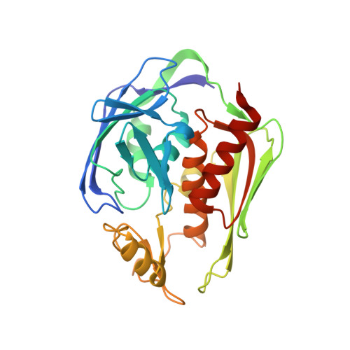
Structure of the deacetylase LpxC bound to the antibiotic CHIR-090: Time-dependent inhibition and specificity in ligand binding

Barb, A.W.Jiang, L.Raetz, C.R.Zhou, P.

(2007) Proc Natl Acad Sci U S A 104: 18433-18438

  • DOI: https://doi.org/10.1073/pnas.0709412104
  • Primary Citation of Related Structures:  
    2JT2

  • PubMed Abstract: 

    The UDP-3-O-(R-3-hydroxyacyl)-N-acetylglucosamine deacetylase LpxC is an essential enzyme of lipid A biosynthesis in Gram-negative bacteria and a promising antibiotic target. CHIR-090, the most potent LpxC inhibitor discovered to date, displays two-step time-dependent inhibition and kills a wide range of Gram-negative pathogens as effectively as ciprofloxacin or tobramycin. In this study, we report the solution structure of the LpxC-CHIR-090 complex. CHIR-090 exploits conserved features of LpxC that are critical for catalysis, including the hydrophobic passage and essential active-site residues. CHIR-090 is adjacent to, but does not occupy, the UDP-binding pocket of LpxC, suggesting that a fragment-based approach may facilitate further optimization of LpxC inhibitors. Additionally, we identified key residues in the Insert II hydrophobic passage that modulate time-dependent inhibition and CHIR-090 resistance. CHIR-090 shares a similar, although previously unrecognized, chemical scaffold with other small-molecule antibiotics such as L-161,240 targeting LpxC, and provides a template for understanding the binding mode of these inhibitors. Consistent with this model, we provide evidence that L-161,240 also occupies the hydrophobic passage.


  • Organizational Affiliation

    Department of Biochemistry, Duke University Medical Center, Durham NC 27710.


Macromolecules
Find similar proteins by:  (by identity cutoff)  |  3D Structure
Entity ID: 1
MoleculeChains Sequence LengthOrganismDetailsImage
UDP-3-O-[3-hydroxymyristoyl] N-acetylglucosamine deacetylase274Aquifex aeolicusMutation(s): 1 
Gene Names: lpxCenvA
EC: 3.5.1
UniProt
Find proteins for O67648 (Aquifex aeolicus (strain VF5))
Explore O67648 
Go to UniProtKB:  O67648
Entity Groups  
Sequence Clusters30% Identity50% Identity70% Identity90% Identity95% Identity100% Identity
UniProt GroupO67648
Sequence Annotations
Expand
  • Reference Sequence
Small Molecules
Ligands 2 Unique
IDChains Name / Formula / InChI Key2D Diagram3D Interactions
C90
Query on C90

Download Ideal Coordinates CCD File 
C [auth A]N-{(1S,2R)-2-hydroxy-1-[(hydroxyamino)carbonyl]propyl}-4-{[4-(morpholin-4-ylmethyl)phenyl]ethynyl}benzamide
C24 H27 N3 O5
FQYBTYFKOHPWQT-VGSWGCGISA-N
ZN
Query on ZN

Download Ideal Coordinates CCD File 
B [auth A]ZINC ION
Zn
PTFCDOFLOPIGGS-UHFFFAOYSA-N
Experimental Data & Validation

Experimental Data

  • Method: SOLUTION NMR
  • Conformers Calculated: 25 
  • Conformers Submitted: 25 
  • Selection Criteria: all calculated structures submitted 

Structure Validation

View Full Validation Report


Entry History 

Deposition Data

Revision History  (Full details and data files)

  • Version 1.0: 2007-12-04
    Type: Initial release
  • Version 1.1: 2011-07-13
    Changes: Version format compliance
  • Version 2.0: 2018-03-14
    Changes: Atomic model, Derived calculations, Experimental preparation
  • Version 2.1: 2021-10-20
    Changes: Database references, Derived calculations