Structure of Pbp-A from Thermosynechococcus Elongatus, a Penicillin-Binding Protein Closely Related to Class a Beta-Lactamases.
Urbach, C., Evrard, C., Pudzaitis, V., Fastrez, J., Soumillion, P., Declercq, J.P.(2009) J Mol Biol 386: 109
- PubMed: 19100272
- DOI: https://doi.org/10.1016/j.jmb.2008.12.001
- Primary Citation of Related Structures:
2J7V, 2J8Y, 2J9O, 2JBF - PubMed Abstract:
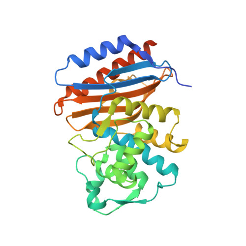
Molecular evolution has always been a subject of discussions, and researchers are interested in understanding how proteins with similar scaffolds can catalyze different reactions. In the superfamily of serine penicillin-recognizing enzymes, D-alanyl-D-alanine peptidases and beta-lactamases are phylogenetically linked but feature large differences of reactivity towards their respective substrates. In particular, while beta-lactamases hydrolyze penicillins very fast, leading to their inactivation, these molecules inhibit d-alanyl-d-alanine peptidases by forming stable covalent penicilloyl enzymes. In cyanobacteria, we have discovered a new family of penicillin-binding proteins (PBPs) presenting all the sequence features of class A beta-lactamases but having a six-amino-acid deletion in the conserved Omega-loop and lacking the essential Glu166 known to be involved in the penicillin hydrolysis mechanism. With the aim of evolving a member of this family into a beta-lactamase, PBP-A from Thermosynechococcus elongatus has been chosen because of its thermostability. Based on sequence alignments, introduction of a glutamate in position 158 of the shorter Omega-loop afforded an enzyme with a 50-fold increase in the rate of penicillin hydrolysis. The crystal structures of PBP-A in the free and penicilloylated forms at 1.9 A resolution and of L158E mutant at 1.5 A resolution were also solved, giving insights in the catalytic mechanism of the proteins. Since all the active-site elements of PBP-A-L158E, including an essential water molecule, are almost perfectly superimposed with those of a class A beta-lactamase such as TEM-1, the question why our mutant is still 5 orders of magnitude less active as a penicillinase remains and our results emphasize how far we are from understanding the secrets of enzymes. Based on the few minor differences between the active sites of PBP-A and TEM-1, mutations were introduced in the L158E enzyme, but while activities on D-Ala-D-Ala mimicking substrates were severely impaired, further improvement in penicillinase activity was unsuccessful.
Organizational Affiliation:
Laboratoire d'Ingénierie des Protéines et des Peptides, Institut des Sciences de la Vie, Université catholique de Louvain, Place Croix du Sud 4-5, bte3, 1348 Louvain la-Neuve, Belgium.