2I94

NMR Structure of recoverin bound to rhodopsin kinase


Experimental Data Snapshot

  • Method: SOLUTION NMR
  • Conformers Calculated: 20 
  • Conformers Submitted: 10 
  • Selection Criteria: structures with the least restraint violations 

wwPDB Validation   3D Report Full Report


This is version 1.3 of the entry. See complete history


Literature

Structural Basis for Calcium-induced Inhibition of Rhodopsin Kinase by Recoverin.

Ames, J.B.Levay, K.Wingard, J.N.Lusin, J.D.Slepak, V.Z.

(2006) J Biol Chem 281: 37237-37245

  • DOI: https://doi.org/10.1074/jbc.M606913200
  • Primary Citation of Related Structures:  
    2I94

  • PubMed Abstract: 

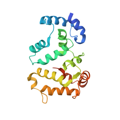
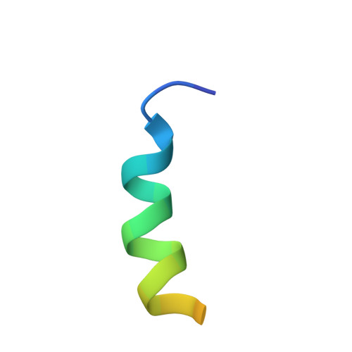
    Recoverin, a member of the neuronal calcium sensor branch of the EF-hand superfamily, serves as a calcium sensor that regulates rhodopsin kinase (RK) activity in retinal rod cells. We report here the NMR structure of Ca(2+)-bound recoverin bound to a functional N-terminal fragment of rhodopsin kinase (residues 1-25, called RK25). The overall main-chain structure of recoverin in the complex is similar to structures of Ca(2+)-bound recoverin in the absence of target (<1.8A root-mean-square deviation). The first eight residues of recoverin at the N terminus are solvent-exposed, enabling the N-terminal myristoyl group to interact with target membranes, and Ca(2+) is bound at the second and third EF-hands of the protein. RK25 in the complex forms an amphipathic helix (residues 4-16). The hydrophobic face of the RK25 helix (Val-9, Val-10, Ala-11, Ala-14, and Phe-15) interacts with an exposed hydrophobic groove on the surface of recoverin lined by side-chain atoms of Trp-31, Phe-35, Phe-49, Ile-52, Tyr-53, Phe-56, Phe-57, Tyr-86, and Leu-90. Residues of recoverin that contact RK25 are highly conserved, suggesting a similar target binding site structure in all neuronal calcium sensor proteins. Site-specific mutagenesis and deletion analysis confirm that the hydrophobic residues at the interface are necessary and sufficient for binding. The recoverin-RK25 complex exhibits Ca(2+)-induced binding to rhodopsin immobilized on concanavalin-A resin. We propose that Ca(2+)-bound recoverin is bound between rhodopsin and RK in a ternary complex on rod outer segment disk membranes, thereby blocking RK interaction with rhodopsin at high Ca(2+).


  • Organizational Affiliation

    Center for Advanced Research in Biotechnology, University of Maryland Biotechnology Institute, Rockville, Maryland 20850, USA. ames@chem.ucdavis.edu


Macromolecules
Find similar proteins by:  (by identity cutoff)  |  3D Structure
Entity ID: 1
MoleculeChains Sequence LengthOrganismDetailsImage
Recoverin202Bos taurusMutation(s): 0 
UniProt
Find proteins for P21457 (Bos taurus)
Explore P21457 
Go to UniProtKB:  P21457
Entity Groups  
Sequence Clusters30% Identity50% Identity70% Identity90% Identity95% Identity100% Identity
UniProt GroupP21457
Sequence Annotations
Expand
  • Reference Sequence

Find similar proteins by:  Sequence   |   3D Structure  

Entity ID: 2
MoleculeChains Sequence LengthOrganismDetailsImage
Rhodopsin kinase25Bos taurusMutation(s): 0 
EC: 2.7.11.14
UniProt
Find proteins for P28327 (Bos taurus)
Explore P28327 
Go to UniProtKB:  P28327
Entity Groups  
Sequence Clusters30% Identity50% Identity70% Identity90% Identity95% Identity100% Identity
UniProt GroupP28327
Sequence Annotations
Expand
  • Reference Sequence
Small Molecules
Ligands 1 Unique
IDChains Name / Formula / InChI Key2D Diagram3D Interactions
CA
Query on CA

Download Ideal Coordinates CCD File 
C [auth A],
D [auth A]
CALCIUM ION
Ca
BHPQYMZQTOCNFJ-UHFFFAOYSA-N
Experimental Data & Validation

Experimental Data

  • Method: SOLUTION NMR
  • Conformers Calculated: 20 
  • Conformers Submitted: 10 
  • Selection Criteria: structures with the least restraint violations 

Structure Validation

View Full Validation Report


Entry History 

Deposition Data

  • Released Date: 2006-10-10 
  • Deposition Author(s): Ames, J.B.

Revision History  (Full details and data files)

  • Version 1.0: 2006-10-10
    Type: Initial release
  • Version 1.1: 2008-05-01
    Changes: Version format compliance
  • Version 1.2: 2011-07-13
    Changes: Version format compliance
  • Version 1.3: 2022-03-09
    Changes: Data collection, Database references, Derived calculations