2I0B

Crystal structure of the GluR6 ligand binding core ELKQ mutant dimer at 1.96 Angstroms Resolution


Experimental Data Snapshot

  • Method: X-RAY DIFFRACTION
  • Resolution: 1.96 Å
  • R-Value Free: 0.211 
  • R-Value Work: 0.172 
  • R-Value Observed: 0.174 

wwPDB Validation   3D Report Full Report


This is version 1.6 of the entry. See complete history


Literature

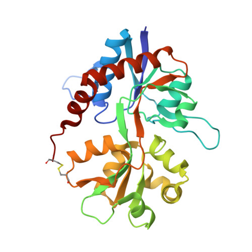
Conformational restriction blocks glutamate receptor desensitization.

Weston, M.C.Schuck, P.Ghosal, A.Rosenmund, C.Mayer, M.L.

(2006) Nat Struct Mol Biol 13: 1120-1127

  • DOI: https://doi.org/10.1038/nsmb1178
  • Primary Citation of Related Structures:  
    2I0B, 2I0C

  • PubMed Abstract: 

    Desensitization is a universal feature of ligand-gated ion channels. Using the crystal structure of the GluR2 L483Y mutant channel as a guide, we attempted to build non-desensitizing kainate-subtype glutamate receptors. Success was achieved for GluR5, GluR6 and GluR7 with intermolecular disulfide cross-links but not by engineering the dimer interface. Crystallographic analysis of the GluR6 Y490C L752C dimer revealed relaxation from the active conformation, which functional studies reveal is not sufficient to trigger desensitization. The equivalent non-desensitizing cross-linked GluR2 mutant retained weak sensitivity to a positive allosteric modulator, which had no effect on GluR2 L483Y. These results establish that the active conformation of AMPA and kainate receptors is conserved and further show that their desensitization requires dimer rearrangements, that subtle structural differences account for their diverse functional properties and that the ligand-binding core dimer is a powerful regulator of ion-channel activity.


  • Organizational Affiliation

    Department of Neuroscience, Baylor College of Medicine, Houston, Texas 77030, USA.


Macromolecules
Find similar proteins by:  (by identity cutoff)  |  3D Structure
Entity ID: 1
MoleculeChains Sequence LengthOrganismDetailsImage
Glutamate receptor, ionotropic kainate 2
A, B, C
259Rattus norvegicusMutation(s): 4 
Gene Names: Grik2Glur6
UniProt
Find proteins for P42260 (Rattus norvegicus)
Explore P42260 
Go to UniProtKB:  P42260
Entity Groups  
Sequence Clusters30% Identity50% Identity70% Identity90% Identity95% Identity100% Identity
UniProt GroupP42260
Sequence Annotations
Expand
  • Reference Sequence
Experimental Data & Validation

Experimental Data

  • Method: X-RAY DIFFRACTION
  • Resolution: 1.96 Å
  • R-Value Free: 0.211 
  • R-Value Work: 0.172 
  • R-Value Observed: 0.174 
  • Space Group: P 43 21 2
Unit Cell:
Length ( Å )Angle ( ˚ )
a = 94.215α = 90
b = 94.215β = 90
c = 219.836γ = 90
Software Package:
Software NamePurpose
REFMACrefinement
SERGUIdata collection
MAR345data collection
HKL-2000data reduction
HKL-2000data scaling
MOLREPphasing

Structure Validation

View Full Validation Report


Entry History 

Deposition Data

  • Released Date: 2006-11-21 
  • Deposition Author(s): Mayer, M.L.

Revision History  (Full details and data files)

  • Version 1.0: 2006-11-21
    Type: Initial release
  • Version 1.1: 2008-05-01
    Changes: Version format compliance
  • Version 1.2: 2011-07-13
    Changes: Advisory, Version format compliance
  • Version 1.3: 2017-08-16
    Changes: Source and taxonomy
  • Version 1.4: 2017-10-18
    Changes: Refinement description
  • Version 1.5: 2021-10-20
    Changes: Database references, Derived calculations
  • Version 1.6: 2024-04-03
    Changes: Data collection, Refinement description