2HR1

Ternary structure of WT M.HhaI C5-Cytosine DNA methyltransferase with unmodified DNA and AdoHcy


Experimental Data Snapshot

  • Method: X-RAY DIFFRACTION
  • Resolution: 1.96 Å
  • R-Value Free: 0.269 
  • R-Value Work: 0.238 
  • R-Value Observed: 0.390 

wwPDB Validation   3D Report Full Report


Ligand Structure Quality Assessment 


This is version 1.3 of the entry. See complete history


Literature

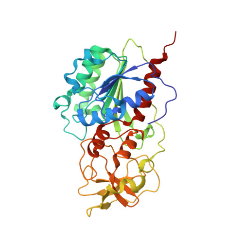
The Role of Arg165 Towards Base Flipping, Base Stabilization and Catalysis in M.HhaI.

Shieh, F.K.Youngblood, B.Reich, N.O.

(2006) J Mol Biol 362: 516-527

  • DOI: https://doi.org/10.1016/j.jmb.2006.07.030
  • Primary Citation of Related Structures:  
    2HR1, 2Z6Q

  • PubMed Abstract: 

    Arg165 forms part of a previously identified base flipping motif in the bacterial DNA cytosine methyltransferase, M.HhaI. Replacement of Arg165 with Ala has no detectable effect on either DNA or AdoMet affinity, yet causes the base flipping and restacking transitions to be decreased approximately 16 and 190-fold respectively, thus confirming the importance of this motif. However, these kinetic changes cannot account for the mutant's observed 10(5)-fold decreased catalytic rate. The mutant enzyme/cognate DNA cocrystal structure (2.79 A resolution) shows the target cytosine to be positioned approximately 30 degrees into the major groove, which is consistent with a major groove pathway for nucleotide flipping. The pyrimidine-sugar chi angle is rotated to approximately +171 degrees, from a range of -95 degrees to -120 degrees in B DNA, and -77 degrees in the WT M.HhaI complex. Thus, Arg165 is important for maintaining the cytosine positioned for nucleophilic attack by Cys81. The cytosine sugar pucker is in the C2'-endo-C3'-exo (South conformation), in contrast to the previously reported C3'-endo (North conformation) described for the original 2.70 A resolution cocrystal structure of the WT M.HhaI/DNA complex. We determined a high resolution structure of the WT M.HhaI/DNA complex (1.96 A) to better determine the sugar pucker. This new structure is similar to the original, lower resolution WT M.HhaI complex, but shows that the sugar pucker is O4'-endo (East conformation), intermediate between the South and North conformers. In summary, Arg165 plays significant roles in base flipping, cytosine positioning, and catalysis. Furthermore, the previously proposed M.HhaI-mediated changes in sugar pucker may not be an important contributor to the base flipping mechanism. These results provide insights into the base flipping and catalytic mechanisms for bacterial and eukaryotic DNA methyltransferases.


  • Organizational Affiliation

    Department of Chemistry and Biochemistry, University of California, Santa Barbara, CA 93106-9510, USA.


Macromolecules

Find similar proteins by:  (by identity cutoff)  |  3D Structure
Entity ID: 3
MoleculeChains Sequence LengthOrganismDetailsImage
Modification methylase HhaIC [auth A]327Haemophilus parahaemolyticusMutation(s): 0 
Gene Names: hhaIM
EC: 2.1.1.73
UniProt
Find proteins for P05102 (Haemophilus parahaemolyticus)
Explore P05102 
Go to UniProtKB:  P05102
Entity Groups  
Sequence Clusters30% Identity50% Identity70% Identity90% Identity95% Identity100% Identity
UniProt GroupP05102
Sequence Annotations
Expand
  • Reference Sequence

Find similar nucleic acids by:  Sequence   |   3D Structure  

Entity ID: 1
MoleculeChains LengthOrganismImage
5'-D(*GP*AP*TP*AP*GP*CP*GP*CP*TP*AP*TP*C)-3'A [auth C]12N/A
Sequence Annotations
Expand
  • Reference Sequence

Find similar nucleic acids by:  Sequence   |   3D Structure  

Entity ID: 2
MoleculeChains LengthOrganismImage
5'-D(*TP*GP*AP*TP*AP*GP*CP*GP*CP*TP*AP*TP*C)-3'B [auth D]13N/A
Sequence Annotations
Expand
  • Reference Sequence
Small Molecules
Ligands 1 Unique
IDChains Name / Formula / InChI Key2D Diagram3D Interactions
SAH
Query on SAH

Download Ideal Coordinates CCD File 
D [auth A]S-ADENOSYL-L-HOMOCYSTEINE
C14 H20 N6 O5 S
ZJUKTBDSGOFHSH-WFMPWKQPSA-N
Experimental Data & Validation

Experimental Data

  • Method: X-RAY DIFFRACTION
  • Resolution: 1.96 Å
  • R-Value Free: 0.269 
  • R-Value Work: 0.238 
  • R-Value Observed: 0.390 
  • Space Group: H 3 2
Unit Cell:
Length ( Å )Angle ( ˚ )
a = 98.72α = 90
b = 98.72β = 90
c = 322.78γ = 120
Software Package:
Software NamePurpose
CNSrefinement
MOSFLMdata reduction
CCP4data scaling
CNSphasing

Structure Validation

View Full Validation Report


Ligand Structure Quality Assessment 


Entry History 

Deposition Data

  • Released Date: 2006-09-19 
  • Deposition Author(s): Shieh, F.

Revision History  (Full details and data files)

  • Version 1.0: 2006-09-19
    Type: Initial release
  • Version 1.1: 2008-05-01
    Changes: Version format compliance
  • Version 1.2: 2011-07-13
    Changes: Version format compliance
  • Version 1.3: 2024-02-14
    Changes: Data collection, Database references, Derived calculations