2HO2

Structure of human FE65-WW domain in complex with hMena peptide.


Experimental Data Snapshot

  • Method: X-RAY DIFFRACTION
  • Resolution: 1.33 Å
  • R-Value Free: 0.214 
  • R-Value Work: 0.172 
  • R-Value Observed: 0.174 

wwPDB Validation   3D Report Full Report


This is version 1.4 of the entry. See complete history


Literature

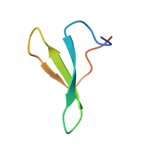
Structural Basis for Polyproline Recognition by the FE65 WW Domain.

Meiyappan, M.Birrane, G.Ladias, J.A.

(2007) J Mol Biol 372: 970-980

  • DOI: https://doi.org/10.1016/j.jmb.2007.06.064
  • Primary Citation of Related Structures:  
    2HO2, 2IDH, 2OEI

  • PubMed Abstract: 

    The neuronal protein FE65 functions in brain development and amyloid precursor protein (APP) signaling through its interaction with the mammalian enabled (Mena) protein and APP, respectively. The recognition of short polyproline sequences in Mena by the FE65 WW domain has a central role in axon guidance and neuronal positioning in the developing brain. We have determined the crystal structures of the human FE65 WW domain (residues 253-289) in the apo form and bound to the peptides PPPPPPLPP and PPPPPPPPPL, which correspond to human Mena residues 313-321 and 347-356, respectively. The FE65 WW domain contains two parallel ligand-binding grooves, XP (formed by residues Y269 and W280) and XP2 (formed by Y269 and W271). Both Mena peptides adopt a polyproline helical II conformation and bind to the WW domain in a forward (N-C) orientation through selection of the PPPPP motif by the XP and XP2 grooves. This mode of ligand recognition is strikingly similar to polyproline interaction with SH3 domains. Importantly, comparison of the FE65 WW structures in the apo and liganded forms shows that the XP2 groove is formed by an induced-fit mechanism that involves movements of the W271 and Y269 side-chains upon ligand binding. These structures elucidate the molecular determinants underlying polyproline ligand selection by the FE65 WW domain and provide a framework for the design of small molecules that would interfere with FE65 WW-ligand interaction and modulate neuronal development and APP signaling.


  • Organizational Affiliation

    Molecular Medicine Laboratory and Macromolecular Crystallography Unit, Division of Experimental Medicine, Harvard Institutes of Medicine, Harvard Medical School, Boston, MA 02115, USA.


Macromolecules
Find similar proteins by:  (by identity cutoff)  |  3D Structure
Entity ID: 1
MoleculeChains Sequence LengthOrganismDetailsImage
Amyloid beta A4 protein-binding family B member 138Homo sapiensMutation(s): 0 
Gene Names: APBB1FE65
UniProt & NIH Common Fund Data Resources
Find proteins for O00213 (Homo sapiens)
Explore O00213 
Go to UniProtKB:  O00213
PHAROS:  O00213
GTEx:  ENSG00000166313 
Entity Groups  
Sequence Clusters30% Identity50% Identity70% Identity90% Identity95% Identity100% Identity
UniProt GroupO00213
Sequence Annotations
Expand
  • Reference Sequence

Find similar proteins by:  Sequence   |   3D Structure  

Entity ID: 2
MoleculeChains Sequence LengthOrganismDetailsImage
Protein enabled homolog10N/AMutation(s): 0 
UniProt & NIH Common Fund Data Resources
Find proteins for Q8N8S7 (Homo sapiens)
Explore Q8N8S7 
Go to UniProtKB:  Q8N8S7
PHAROS:  Q8N8S7
GTEx:  ENSG00000154380 
Entity Groups  
Sequence Clusters30% Identity50% Identity70% Identity90% Identity95% Identity100% Identity
UniProt GroupQ8N8S7
Sequence Annotations
Expand
  • Reference Sequence
Small Molecules
Ligands 1 Unique
IDChains Name / Formula / InChI Key2D Diagram3D Interactions
GOL
Query on GOL

Download Ideal Coordinates CCD File 
C [auth A]GLYCEROL
C3 H8 O3
PEDCQBHIVMGVHV-UHFFFAOYSA-N
Experimental Data & Validation

Experimental Data

  • Method: X-RAY DIFFRACTION
  • Resolution: 1.33 Å
  • R-Value Free: 0.214 
  • R-Value Work: 0.172 
  • R-Value Observed: 0.174 
  • Space Group: P 31 2 1
Unit Cell:
Length ( Å )Angle ( ˚ )
a = 41.661α = 90
b = 41.661β = 90
c = 38.633γ = 120
Software Package:
Software NamePurpose
DENZOdata reduction
SCALEPACKdata scaling
REFMACrefinement
PDB_EXTRACTdata extraction
HKL-2000data reduction
HKL-2000data scaling
MOLREPphasing

Structure Validation

View Full Validation Report


Entry History 

Deposition Data

Revision History  (Full details and data files)

  • Version 1.0: 2007-07-10
    Type: Initial release
  • Version 1.1: 2007-10-16
    Changes: Version format compliance
  • Version 1.2: 2011-07-13
    Changes: Non-polymer description, Version format compliance
  • Version 1.3: 2017-10-18
    Changes: Refinement description
  • Version 1.4: 2024-02-14
    Changes: Data collection, Database references, Derived calculations