2HIB

human formylglycine generating enzyme, C336S mutant, iodide co-crystallization


Experimental Data Snapshot

  • Method: X-RAY DIFFRACTION
  • Resolution: 2.00 Å
  • R-Value Free: 0.238 
  • R-Value Work: 0.172 
  • R-Value Observed: 0.175 

wwPDB Validation   3D Report Full Report


This is version 2.2 of the entry. See complete history


Literature

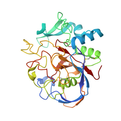
Probing the oxygen-binding site of the human formylglycine-generating enzyme using halide ions.

Roeser, D.Schmidt, B.Preusser-Kunze, A.Rudolph, M.G.

(2007) Acta Crystallogr D Biol Crystallogr 63: 621-627

  • DOI: https://doi.org/10.1107/S0907444907009961
  • Primary Citation of Related Structures:  
    2HI8, 2HIB

  • PubMed Abstract: 

    The catalytic residue in sulfatases is a unique formylglycine that is post-translationally generated by oxidation of a cysteine or serine precursor. Molecular oxygen oxidizes the cysteine precursor in eukaryotic sulfatases, a reaction that is catalysed by the formylglycine-generating enzyme FGE. Previously, FGE was crystallized in complex with a chloride ion which, based on its similar polarizability and hydrophobicity, indicates the site of molecular oxygen binding. Here, two structures of FGE in complex with bromide and iodide were determined in order to further delineate the volume and stereochemical restraints of the oxygen-binding site for potential reaction intermediates. Anomalous difference density maps unambiguously assigned the nature of the halide ions. Unexpectedly, data collected at a wavelength of 1.54 A from the iodide-containing crystal and data collected at a wavelength of 0.8 A from a bromide-containing crystal were sufficient for SIRAS phasing.


  • Organizational Affiliation

    Department of Molecular Structural Biology, University of Göttingen, D-37077 Göttingen, Germany.


Macromolecules
Find similar proteins by:  (by identity cutoff)  |  3D Structure
Entity ID: 1
MoleculeChains Sequence LengthOrganismDetailsImage
Sulfatase-modifying factor 1A [auth X]286Homo sapiensMutation(s): 1 
UniProt & NIH Common Fund Data Resources
Find proteins for Q8NBK3 (Homo sapiens)
Explore Q8NBK3 
Go to UniProtKB:  Q8NBK3
PHAROS:  Q8NBK3
GTEx:  ENSG00000144455 
Entity Groups  
Sequence Clusters30% Identity50% Identity70% Identity90% Identity95% Identity100% Identity
UniProt GroupQ8NBK3
Sequence Annotations
Expand
  • Reference Sequence
Oligosaccharides

Help

Entity ID: 2
MoleculeChains Length2D Diagram Glycosylation3D Interactions
2-acetamido-2-deoxy-beta-D-glucopyranose-(1-4)-2-acetamido-2-deoxy-beta-D-glucopyranoseB [auth A]2N-Glycosylation
Glycosylation Resources
GlyTouCan:  G42666HT
GlyCosmos:  G42666HT
GlyGen:  G42666HT
Small Molecules
Ligands 2 Unique
IDChains Name / Formula / InChI Key2D Diagram3D Interactions
IOD
Query on IOD

Download Ideal Coordinates CCD File 
E [auth X]
F [auth X]
G [auth X]
H [auth X]
I [auth X]
E [auth X],
F [auth X],
G [auth X],
H [auth X],
I [auth X],
J [auth X],
K [auth X],
L [auth X],
M [auth X],
N [auth X],
O [auth X]
IODIDE ION
I
XMBWDFGMSWQBCA-UHFFFAOYSA-M
CA
Query on CA

Download Ideal Coordinates CCD File 
C [auth X],
D [auth X]
CALCIUM ION
Ca
BHPQYMZQTOCNFJ-UHFFFAOYSA-N
Experimental Data & Validation

Experimental Data

  • Method: X-RAY DIFFRACTION
  • Resolution: 2.00 Å
  • R-Value Free: 0.238 
  • R-Value Work: 0.172 
  • R-Value Observed: 0.175 
  • Space Group: P 21 21 2
Unit Cell:
Length ( Å )Angle ( ˚ )
a = 61.89α = 90
b = 109.11β = 90
c = 43.492γ = 90
Software Package:
Software NamePurpose
REFMACrefinement
HKL-2000data reduction
HKL-2000data scaling

Structure Validation

View Full Validation Report


Entry History 

Deposition Data

Revision History  (Full details and data files)

  • Version 1.0: 2007-05-01
    Type: Initial release
  • Version 1.1: 2008-05-01
    Changes: Version format compliance
  • Version 1.2: 2011-07-13
    Changes: Non-polymer description, Version format compliance
  • Version 2.0: 2020-07-29
    Type: Remediation
    Reason: Carbohydrate remediation
    Changes: Atomic model, Data collection, Derived calculations, Structure summary
  • Version 2.1: 2021-11-10
    Changes: Database references, Structure summary
  • Version 2.2: 2023-10-25
    Changes: Data collection, Refinement description