2HEA

CONTRIBUTION OF WATER MOLECULES IN THE INTERIOR OF A PROTEIN TO THE CONFORMATIONAL STABILITY


Experimental Data Snapshot

  • Method: X-RAY DIFFRACTION
  • Resolution: 1.80 Å
  • R-Value Work: 0.156 
  • R-Value Observed: 0.156 

wwPDB Validation   3D Report Full Report


This is version 1.4 of the entry. See complete history


Literature

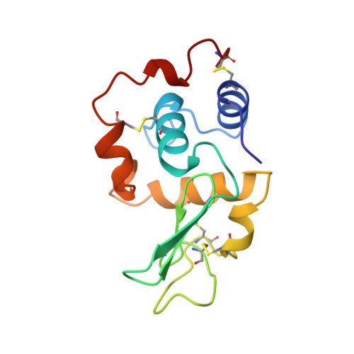
Contribution of water molecules in the interior of a protein to the conformational stability.

Takano, K.Funahashi, J.Yamagata, Y.Fujii, S.Yutani, K.

(1997) J Mol Biol 274: 132-142

  • DOI: https://doi.org/10.1006/jmbi.1997.1365
  • Primary Citation of Related Structures:  
    2HEA, 2HEB, 2HEC, 2HED, 2HEE, 2HEF

  • PubMed Abstract: 

    Water molecules frequently occur in the interior of globular proteins. To elucidate the contribution of buried water molecules to the conformational stability of a protein, we examined the crystal structures and the thermodynamic parameters of denaturation of six Ile to Ala/Gly mutant human lysozymes, in which a cavity is created at each mutation site by the substitution of a smaller side-chain for a larger one. One or two ordered water molecules were found in the cavities created in some mutants (I106A, I59A and I59G). The cavity volumes for these three mutants were bigger than those that remained empty in the other mutants. The stability of the mutant proteins with the newly introduced water molecules was about 8 kJ/mol higher than that expected from the change in hydrophobic surface area (DeltaDeltaASAHP) exposed upon denaturation. It was concluded that a water molecule in a cavity created in the interior of a protein contributes favorably to the stability.


  • Organizational Affiliation

    Institute for Protein Research, Osaka University, Japan.


Macromolecules
Find similar proteins by:  (by identity cutoff)  |  3D Structure
Entity ID: 1
MoleculeChains Sequence LengthOrganismDetailsImage
LYSOZYME130Homo sapiensMutation(s): 1 
Gene Names: HUMAN LYSOZYME WITH ILE 106
EC: 3.2.1.17
UniProt & NIH Common Fund Data Resources
Find proteins for P61626 (Homo sapiens)
Explore P61626 
Go to UniProtKB:  P61626
PHAROS:  P61626
GTEx:  ENSG00000090382 
Entity Groups  
Sequence Clusters30% Identity50% Identity70% Identity90% Identity95% Identity100% Identity
UniProt GroupP61626
Sequence Annotations
Expand
  • Reference Sequence
Small Molecules
Ligands 1 Unique
IDChains Name / Formula / InChI Key2D Diagram3D Interactions
NA
Query on NA

Download Ideal Coordinates CCD File 
B [auth A]SODIUM ION
Na
FKNQFGJONOIPTF-UHFFFAOYSA-N
Experimental Data & Validation

Experimental Data

  • Method: X-RAY DIFFRACTION
  • Resolution: 1.80 Å
  • R-Value Work: 0.156 
  • R-Value Observed: 0.156 
  • Space Group: P 21 21 21
Unit Cell:
Length ( Å )Angle ( ˚ )
a = 56.99α = 90
b = 60.9β = 90
c = 33.77γ = 90
Software Package:
Software NamePurpose
PROCESSdata collection
X-PLORmodel building
X-PLORrefinement
PROCESSdata reduction
X-PLORphasing

Structure Validation

View Full Validation Report


Entry History 

Deposition Data

Revision History  (Full details and data files)

  • Version 1.0: 1998-01-14
    Type: Initial release
  • Version 1.1: 2008-03-24
    Changes: Version format compliance
  • Version 1.2: 2011-07-13
    Changes: Version format compliance
  • Version 1.3: 2017-11-29
    Changes: Derived calculations, Other
  • Version 1.4: 2021-11-03
    Changes: Database references, Derived calculations