2GZ1

Structure of Aspartate Semialdehyde Dehydrogenase (ASADH) from Streptococcus pneumoniae complexed with NADP


Experimental Data Snapshot

  • Method: X-RAY DIFFRACTION
  • Resolution: 1.80 Å
  • R-Value Free: 0.191 
  • R-Value Work: 0.160 
  • R-Value Observed: 0.162 

wwPDB Validation   3D Report Full Report


Ligand Structure Quality Assessment 


This is version 1.3 of the entry. See complete history


Literature

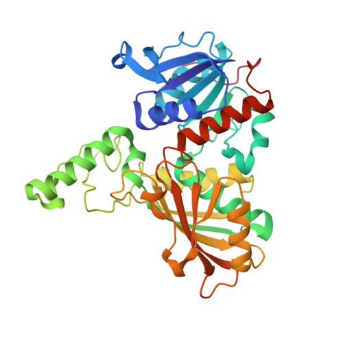
Examination of key intermediates in the catalytic cycle of aspartate-beta-semialdehyde dehydrogenase from a gram-positive infectious bacteria.

Faehnle, C.R.Le Coq, J.Liu, X.Viola, R.E.

(2006) J Biol Chem 281: 31031-31040

  • DOI: https://doi.org/10.1074/jbc.M605926200
  • Primary Citation of Related Structures:  
    2GYY, 2GZ1, 2GZ2, 2GZ3

  • PubMed Abstract: 

    Aspartate-beta-semialdehyde dehydrogenase (ASADH) catalyzes a critical branch point transformation in amino acid bio-synthesis. The products of the aspartate pathway are essential in microorganisms, and this entire pathway is absent in mammals, making this enzyme an attractive target for antibiotic development. The first structure of an ASADH from a Gram-positive bacterium, Streptococcus pneumoniae, has now been determined. The overall structure of the apoenzyme has a similar fold to those of the Gram-negative and archaeal ASADHs but contains some interesting structural variations that can be exploited for inhibitor design. Binding of the coenzyme NADP, as well as a truncated nucleotide analogue, into an alternative conformation from that observed in Gram-negative ASADHs causes an enzyme domain closure that precedes catalysis. The covalent acyl-enzyme intermediate was trapped by soaking the substrate into crystals of the coenzyme complex, and the structure of this elusive intermediate provides detailed insights into the catalytic mechanism.


  • Organizational Affiliation

    Department of Chemistry, University of Toledo, Toledo, Ohio 43606, USA.


Macromolecules
Find similar proteins by:  (by identity cutoff)  |  3D Structure
Entity ID: 1
MoleculeChains Sequence LengthOrganismDetailsImage
Aspartate beta-semialdehyde dehydrogenase
A, B
366Streptococcus pneumoniaeMutation(s): 0 
Gene Names: asd
EC: 1.2.1.11
UniProt
Find proteins for A0A0H2UPS5 (Streptococcus pneumoniae serotype 4 (strain ATCC BAA-334 / TIGR4))
Explore A0A0H2UPS5 
Go to UniProtKB:  A0A0H2UPS5
Entity Groups  
Sequence Clusters30% Identity50% Identity70% Identity90% Identity95% Identity100% Identity
UniProt GroupA0A0H2UPS5
Sequence Annotations
Expand
  • Reference Sequence
Small Molecules
Ligands 1 Unique
IDChains Name / Formula / InChI Key2D Diagram3D Interactions
NAP
Query on NAP

Download Ideal Coordinates CCD File 
C [auth A],
D [auth B]
NADP NICOTINAMIDE-ADENINE-DINUCLEOTIDE PHOSPHATE
C21 H28 N7 O17 P3
XJLXINKUBYWONI-NNYOXOHSSA-N
Binding Affinity Annotations 
IDSourceBinding Affinity
NAP Binding MOAD:  2GZ1 Kd: 6000 (nM) from 1 assay(s)
Experimental Data & Validation

Experimental Data

  • Method: X-RAY DIFFRACTION
  • Resolution: 1.80 Å
  • R-Value Free: 0.191 
  • R-Value Work: 0.160 
  • R-Value Observed: 0.162 
  • Space Group: P 1 21 1
Unit Cell:
Length ( Å )Angle ( ˚ )
a = 59.709α = 90
b = 99.165β = 100.83
c = 64.525γ = 90
Software Package:
Software NamePurpose
d*TREKdata scaling
PHASERphasing
REFMACrefinement
PDB_EXTRACTdata extraction
CrystalCleardata reduction

Structure Validation

View Full Validation Report


Ligand Structure Quality Assessment 


Entry History 

Deposition Data

Revision History  (Full details and data files)

  • Version 1.0: 2006-08-15
    Type: Initial release
  • Version 1.1: 2008-02-06
    Changes: Version format compliance
  • Version 1.2: 2011-07-13
    Changes: Derived calculations, Version format compliance
  • Version 1.3: 2023-08-30
    Changes: Data collection, Database references, Derived calculations, Refinement description