2GMU

Crystal structure of E coli GDP-4-keto-6-deoxy-D-mannose-3-dehydratase complexed with PLP-glutamate ketimine intermediate


Experimental Data Snapshot

  • Method: X-RAY DIFFRACTION
  • Resolution: 1.90 Å
  • R-Value Free: 0.231 
  • R-Value Work: 0.174 
  • R-Value Observed: 0.176 

wwPDB Validation   3D Report Full Report


Ligand Structure Quality Assessment 


This is version 1.4 of the entry. See complete history


Literature

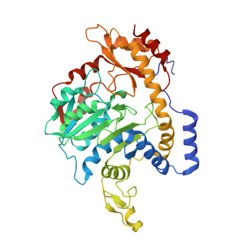
The structure of GDP-4-keto-6-deoxy-D-mannose-3-dehydratase: a unique coenzyme B6-dependent enzyme.

Cook, P.D.Thoden, J.B.Holden, H.M.

(2006) Protein Sci 15: 2093-2106

  • DOI: https://doi.org/10.1110/ps.062328306
  • Primary Citation of Related Structures:  
    2GMS, 2GMU

  • PubMed Abstract: 

    L-colitose is a 3,6-dideoxysugar found in the O-antigens of some Gram-negative bacteria such as Escherichia coli and in marine bacteria such as Pseudoalteromonas tetraodonis. The focus of this investigation, GDP-4-keto-6-deoxy-D-mannose-3-dehydratase, catalyzes the third step in colitose production, which is the removal of the hydroxyl group at C3' of GDP-4-keto-6-deoxymannose. It is an especially intriguing PLP-dependent enzyme in that it acts as both a transaminase and a dehydratase. Here we present the first X-ray structure of this enzyme isolated from E. coli Strain 5a, type O55:H7. The two subunits of the protein form a tight dimer with a buried surface area of approximately 5000 A2. This is a characteristic feature of the aspartate aminotransferase superfamily. Although the PLP-binding pocket is formed primarily by one subunit, there is a loop, delineated by Phe 240 to Glu 253 in the second subunit, that completes the active site architecture. The hydrated form of PLP was observed in one of the enzyme/cofactor complexes described here. Amino acid residues involved in anchoring the cofactor to the protein include Gly 56, Ser 57, Asp 159, Glu 162, and Ser 183 from one subunit and Asn 248 from the second monomer. In the second enzyme/cofactor complex reported, a glutamate ketimine intermediate was found trapped in the active site. Taken together, these two structures, along with previously reported biochemical data, support the role of His 188 as the active site base required for catalysis.


  • Organizational Affiliation

    Department of Biochemistry, University of Wisconsin-Madison, Wisconsin 53706, USA.


Macromolecules
Find similar proteins by:  (by identity cutoff)  |  3D Structure
Entity ID: 1
MoleculeChains Sequence LengthOrganismDetailsImage
Putative pyridoxamine 5-phosphate-dependent dehydrase, WbdK
A, B
390Escherichia coli O55:H7Mutation(s): 0 
Gene Names: wbdK
UniProt
Find proteins for Q9F118 (Escherichia coli)
Explore Q9F118 
Go to UniProtKB:  Q9F118
Entity Groups  
Sequence Clusters30% Identity50% Identity70% Identity90% Identity95% Identity100% Identity
UniProt GroupQ9F118
Sequence Annotations
Expand
  • Reference Sequence
Small Molecules
Ligands 2 Unique
IDChains Name / Formula / InChI Key2D Diagram3D Interactions
PDG
Query on PDG

Download Ideal Coordinates CCD File 
D [auth A],
E [auth B]
N-({3-HYDROXY-2-METHYL-5-[(PHOSPHONOOXY)METHYL]PYRIDIN-4-YL}METHYL)-D-GLUTAMIC ACID
C13 H19 N2 O9 P
JMRKOGDJNHPMHS-SNVBAGLBSA-N
MG
Query on MG

Download Ideal Coordinates CCD File 
C [auth A]MAGNESIUM ION
Mg
JLVVSXFLKOJNIY-UHFFFAOYSA-N
Experimental Data & Validation

Experimental Data

  • Method: X-RAY DIFFRACTION
  • Resolution: 1.90 Å
  • R-Value Free: 0.231 
  • R-Value Work: 0.174 
  • R-Value Observed: 0.176 
  • Space Group: P 21 21 21
Unit Cell:
Length ( Å )Angle ( ˚ )
a = 74.9α = 90
b = 88β = 90
c = 126γ = 90
Software Package:
Software NamePurpose
FRAMBOdata collection
XDSdata reduction
PHASERphasing
TNTrefinement
XDSdata scaling

Structure Validation

View Full Validation Report


Ligand Structure Quality Assessment 


Entry History 

Deposition Data

Revision History  (Full details and data files)

  • Version 1.0: 2006-09-05
    Type: Initial release
  • Version 1.1: 2008-05-01
    Changes: Version format compliance
  • Version 1.2: 2011-07-13
    Changes: Version format compliance
  • Version 1.3: 2017-10-18
    Changes: Refinement description
  • Version 1.4: 2023-08-30
    Changes: Data collection, Database references, Derived calculations, Refinement description