Crystal Structure of Mammalian Cysteine Dioxygenase: A NOVEL MONONUCLEAR IRON CENTER FOR CYSTEINE THIOL OXIDATION.
Simmons, C.R., Liu, Q., Huang, Q., Hao, Q., Begley, T.P., Karplus, P.A., Stipanuk, M.H.(2006) J Biol Chem 281: 18723-18733
- PubMed: 16611640
- DOI: https://doi.org/10.1074/jbc.M601555200
- Primary Citation of Related Structures:
2B5H, 2GH2 - PubMed Abstract:
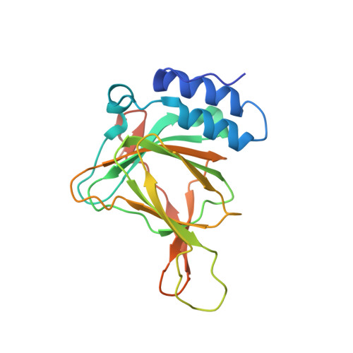
Cysteine dioxygenase is a mononuclear iron-dependent enzyme responsible for the oxidation of cysteine with molecular oxygen to form cysteine sulfinate. This reaction commits cysteine to either catabolism to sulfate and pyruvate or the taurine biosynthetic pathway. Cysteine dioxygenase is a member of the cupin superfamily of proteins. The crystal structure of recombinant rat cysteine dioxygenase has been determined to 1.5-A resolution, and these results confirm the canonical cupin beta-sandwich fold and the rare cysteinyltyrosine intramolecular cross-link (between Cys(93) and Tyr(157)) seen in the recently reported murine cysteine dioxygenase structure. In contrast to the catalytically inactive mononuclear Ni(II) metallocenter present in the murine structure, crystallization of a catalytically competent preparation of rat cysteine dioxygenase revealed a novel tetrahedrally coordinated mononuclear iron center involving three histidines (His(86), His(88), and His(140)) and a water molecule. Attempts to acquire a structure with bound ligand using either cocrystallization or soaking crystals with cysteine revealed the formation of a mixed disulfide involving Cys(164) near the active site, which may explain previously observed substrate inhibition. This work provides a framework for understanding the molecular mechanisms involved in thiol dioxygenation and sets the stage for exploration of the chemistry of both the novel mononuclear iron center and the catalytic role of the cysteinyl-tyrosine linkage.
Organizational Affiliation:
Division of Nutritional Sciences, Cornell University, Ithaca, NY 14853-8001, USA.