2G1M

Cellular Oxygen Sensing: Crystal Structure of Hypoxia-Inducible Factor Prolyl Hydroxylase (PHD2)


Experimental Data Snapshot

  • Method: X-RAY DIFFRACTION
  • Resolution: 2.20 Å
  • R-Value Free: 0.289 
  • R-Value Work: 0.216 
  • R-Value Observed: 0.216 

wwPDB Validation   3D Report Full Report


Ligand Structure Quality Assessment 


This is version 1.4 of the entry. See complete history


Literature

Cellular oxygen sensing: Crystal structure of hypoxia-inducible factor prolyl hydroxylase (PHD2).

McDonough, M.A.Li, V.Flashman, E.Chowdhury, R.Mohr, C.Lienard, B.M.Zondlo, J.Oldham, N.J.Clifton, I.J.Lewis, J.McNeill, L.A.Kurzeja, R.J.Hewitson, K.S.Yang, E.Jordan, S.Syed, R.S.Schofield, C.J.

(2006) Proc Natl Acad Sci U S A 103: 9814-9819

  • DOI: https://doi.org/10.1073/pnas.0601283103
  • Primary Citation of Related Structures:  
    2G19, 2G1M

  • PubMed Abstract: 

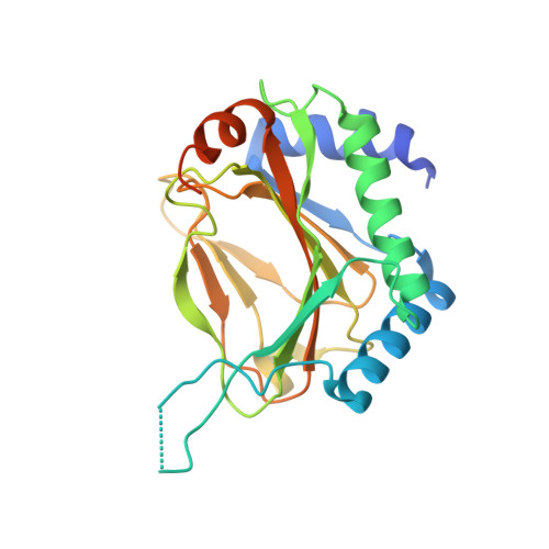
    Cellular and physiological responses to changes in dioxygen levels in metazoans are mediated via the posttranslational oxidation of hypoxia-inducible transcription factor (HIF). Hydroxylation of conserved prolyl residues in the HIF-alpha subunit, catalyzed by HIF prolyl-hydroxylases (PHDs), signals for its proteasomal degradation. The requirement of the PHDs for dioxygen links changes in dioxygen levels with the transcriptional regulation of the gene array that enables the cellular response to chronic hypoxia; the PHDs thus act as an oxygen-sensing component of the HIF system, and their inhibition mimics the hypoxic response. We describe crystal structures of the catalytic domain of human PHD2, an important prolyl-4-hydroxylase in the human hypoxic response in normal cells, in complex with Fe(II) and an inhibitor to 1.7 A resolution. PHD2 crystallizes as a homotrimer and contains a double-stranded beta-helix core fold common to the Fe(II) and 2-oxoglutarate-dependant dioxygenase family, the residues of which are well conserved in the three human PHD enzymes (PHD 1-3). The structure provides insights into the hypoxic response, helps to rationalize a clinically observed mutation leading to familial erythrocytosis, and will aid in the design of PHD selective inhibitors for the treatment of anemia and ischemic disease.


  • Organizational Affiliation

    Oxford Centre for Molecular Sciences and Department of Chemistry, University of Oxford, Mansfield Road, Oxford OX1 3TA, United Kingdom.


Macromolecules
Find similar proteins by:  (by identity cutoff)  |  3D Structure
Entity ID: 1
MoleculeChains Sequence LengthOrganismDetailsImage
Egl nine homolog 1246Homo sapiensMutation(s): 0 
Gene Names: EGLN1C1orf12
EC: 1.14.11
UniProt & NIH Common Fund Data Resources
Find proteins for Q9GZT9 (Homo sapiens)
Explore Q9GZT9 
Go to UniProtKB:  Q9GZT9
PHAROS:  Q9GZT9
GTEx:  ENSG00000135766 
Entity Groups  
Sequence Clusters30% Identity50% Identity70% Identity90% Identity95% Identity100% Identity
UniProt GroupQ9GZT9
Sequence Annotations
Expand
  • Reference Sequence
Small Molecules
Ligands 2 Unique
IDChains Name / Formula / InChI Key2D Diagram3D Interactions
4HG
Query on 4HG

Download Ideal Coordinates CCD File 
C [auth A]N-[(4-HYDROXY-8-IODOISOQUINOLIN-3-YL)CARBONYL]GLYCINE
C12 H9 I N2 O4
FUMNLXHPILGSLC-UHFFFAOYSA-N
FE2
Query on FE2

Download Ideal Coordinates CCD File 
B [auth A]FE (II) ION
Fe
CWYNVVGOOAEACU-UHFFFAOYSA-N
Binding Affinity Annotations 
IDSourceBinding Affinity
4HG BindingDB:  2G1M IC50: 1400 (nM) from 1 assay(s)
Experimental Data & Validation

Experimental Data

  • Method: X-RAY DIFFRACTION
  • Resolution: 2.20 Å
  • R-Value Free: 0.289 
  • R-Value Work: 0.216 
  • R-Value Observed: 0.216 
  • Space Group: P 63
Unit Cell:
Length ( Å )Angle ( ˚ )
a = 110.742α = 90
b = 110.742β = 90
c = 39.986γ = 120
Software Package:
Software NamePurpose
SAINTdata scaling
SADABSdata reduction
SOLVEphasing
RESOLVEmodel building
CNSrefinement
SAINTdata reduction
SADABSdata scaling

Structure Validation

View Full Validation Report


Ligand Structure Quality Assessment 


Entry History 

Deposition Data

Revision History  (Full details and data files)

  • Version 1.0: 2006-06-13
    Type: Initial release
  • Version 1.1: 2008-05-01
    Changes: Version format compliance
  • Version 1.2: 2011-07-13
    Changes: Version format compliance
  • Version 1.3: 2017-10-18
    Changes: Refinement description
  • Version 1.4: 2024-02-14
    Changes: Data collection, Database references, Derived calculations