2FZ2

Structure of Turnip Yellow Mosaic Virus at 100 K


Experimental Data Snapshot

  • Method: X-RAY DIFFRACTION
  • Resolution: 2.90 Å
  • R-Value Free: 0.289 
  • R-Value Work: 0.276 
  • R-Value Observed: 0.277 

wwPDB Validation   3D Report Full Report


This is version 1.3 of the entry. See complete history


Literature

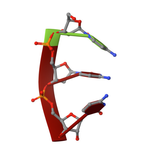
The RNA of turnip yellow mosaic virus exhibits icosahedral order.

Larson, S.B.Lucas, R.W.Greenwood, A.McPherson, A.

(2005) Virology 334: 245-254

  • DOI: https://doi.org/10.1016/j.virol.2005.01.036
  • Primary Citation of Related Structures:  
    2FZ1, 2FZ2

  • PubMed Abstract: 

    Difference electron density maps, based on structure factor amplitudes and experimental phases from crystals of wild-type turnip yellow mosaic virus and those of empty capsids prepared by freeze-thawing, show a large portion of the encapsidated RNA to have an icosahedral distribution. Four unique segments of base-paired, double-helical RNA, one to two turns in length, lie between 33-A and 101-A radius and are organized about either 2-fold or 5-fold icosahedral axes. In addition, single-stranded loops of RNA invade the pentameric and hexameric capsomeres where they contact the interior capsid surface. The remaining RNA, not seen in electron density maps, must serve as connecting links between these secondary structural elements and is likely icosahedrally disordered. The distribution of RNA observed crystallographically appears to be in agreement with models based on biochemical data and secondary structural analyses.


  • Organizational Affiliation

    Department of Molecular Biology and Biochemistry, University of California, Irvine, CA 92697-3900, USA.


Macromolecules

Find similar proteins by:  (by identity cutoff)  |  3D Structure
Entity ID: 2
MoleculeChains Sequence LengthOrganismDetailsImage
Coat proteinB [auth A],
C [auth B],
D [auth C]
189Turnip yellow mosaic virus (Australian isolate)Mutation(s): 3 
UniProt
Find proteins for P20125 (Turnip yellow mosaic virus (isolate Australia))
Explore P20125 
Go to UniProtKB:  P20125
Entity Groups  
Sequence Clusters30% Identity50% Identity70% Identity90% Identity95% Identity100% Identity
UniProt GroupP20125
Sequence Annotations
Expand
  • Reference Sequence

Find similar nucleic acids by:  Sequence   |   3D Structure  

Entity ID: 1
MoleculeChains LengthOrganismImage
5'-R(*CP*CP*C)-3'A [auth D]3N/A
Sequence Annotations
Expand
  • Reference Sequence
Experimental Data & Validation

Experimental Data

  • Method: X-RAY DIFFRACTION
  • Resolution: 2.90 Å
  • R-Value Free: 0.289 
  • R-Value Work: 0.276 
  • R-Value Observed: 0.277 
  • Space Group: P 64 2 2
Unit Cell:
Length ( Å )Angle ( ˚ )
a = 511.53α = 90
b = 511.53β = 90
c = 303.76γ = 120
Software Package:
Software NamePurpose
CNSrefinement
DENZOdata reduction
SCALEPACKdata scaling
CNSphasing

Structure Validation

View Full Validation Report


Entry History 

Deposition Data

Revision History  (Full details and data files)

  • Version 1.0: 2006-02-21
    Type: Initial release
  • Version 1.1: 2008-05-01
    Changes: Version format compliance
  • Version 1.2: 2011-07-13
    Changes: Source and taxonomy, Version format compliance
  • Version 1.3: 2023-08-30
    Changes: Data collection, Database references, Derived calculations, Refinement description