2FSD

A Common Fold for the Receptor Binding Domains of Lactococcal Phages? The Crystal Structure of the Head Domain of Phage bIL170


Experimental Data Snapshot

  • Method: X-RAY DIFFRACTION
  • Resolution: 2.30 Å
  • R-Value Free: 0.268 
  • R-Value Work: 0.242 
  • R-Value Observed: 0.243 

wwPDB Validation   3D Report Full Report


This is version 1.3 of the entry. See complete history


Literature

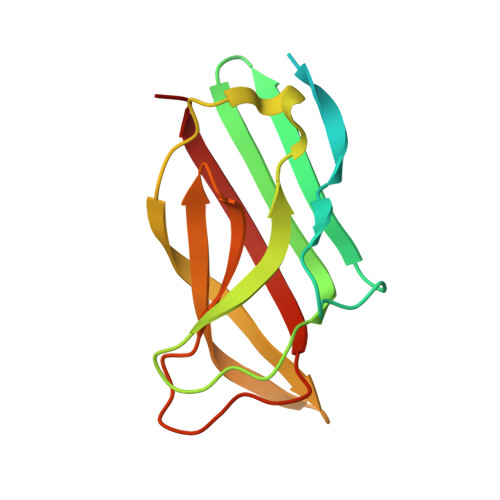
Crystal Structure of the Receptor-Binding Protein Head Domain from Lactococcus lactis Phage bIL170

Ricagno, S.Campanacci, V.Blangy, S.Spinelli, S.Tremblay, D.Moineau, S.Tegoni, M.Cambillau, C.

(2006) J Virol 80: 9331-9335

  • DOI: https://doi.org/10.1128/JVI.01160-06
  • Primary Citation of Related Structures:  
    2FSD

  • PubMed Abstract: 

    Lactococcus lactis, a gram-positive bacterium widely used by the dairy industry, is subject to lytic phage infections. In the first step of infection, phages recognize the host saccharidic receptor using their receptor binding protein (RBP). Here, we report the 2.30-A-resolution crystal structure of the RBP head domain from phage bIL170. The structure of the head monomer is remarkably close to those of other lactococcal phages, p2 and TP901-1, despite any sequence identity with them. The knowledge of the three-dimensional structures of three RBPs gives a better insight into the module exchanges which have occurred among phages.


  • Organizational Affiliation

    Architecture et Fonction des Macromolécules Biologiques, UMR 6098 CNRS and Universités Aix-Marseille I & II, Campus de Luminy, Case 932, 13288 Marseille Cedex 09, France.


Macromolecules
Find similar proteins by:  (by identity cutoff)  |  3D Structure
Entity ID: 1
MoleculeChains Sequence LengthOrganismDetailsImage
putative baseplate protein
A, B
142Bacteriophage sp.Mutation(s): 0 
UniProt
Find proteins for O80120 (Skunavirus bIL170)
Explore O80120 
Go to UniProtKB:  O80120
Entity Groups  
Sequence Clusters30% Identity50% Identity70% Identity90% Identity95% Identity100% Identity
UniProt GroupO80120
Sequence Annotations
Expand
  • Reference Sequence
Experimental Data & Validation

Experimental Data

  • Method: X-RAY DIFFRACTION
  • Resolution: 2.30 Å
  • R-Value Free: 0.268 
  • R-Value Work: 0.242 
  • R-Value Observed: 0.243 
  • Space Group: P 31 2 1
Unit Cell:
Length ( Å )Angle ( ˚ )
a = 69.65α = 90
b = 69.65β = 90
c = 95.356γ = 120
Software Package:
Software NamePurpose
REFMACrefinement
MOSFLMdata reduction
CCP4data scaling
SHELXSphasing

Structure Validation

View Full Validation Report


Entry History 

Deposition Data

Revision History  (Full details and data files)

  • Version 1.0: 2006-09-05
    Type: Initial release
  • Version 1.1: 2008-05-01
    Changes: Version format compliance
  • Version 1.2: 2011-07-13
    Changes: Version format compliance
  • Version 1.3: 2024-03-13
    Changes: Data collection, Database references