2F89

Crystal structure of human FPPS in complex with pamidronate


Experimental Data Snapshot

  • Method: X-RAY DIFFRACTION
  • Resolution: 2.60 Å
  • R-Value Free: 0.267 
  • R-Value Work: 0.199 
  • R-Value Observed: 0.206 

wwPDB Validation   3D Report Full Report


Ligand Structure Quality Assessment 


This is version 1.4 of the entry. See complete history


Literature

Structural basis for the exceptional in vivo efficacy of bisphosphonate drugs.

Rondeau, J.M.Bitsch, F.Bourgier, E.Geiser, M.Hemmig, R.Kroemer, M.Lehmann, S.Ramage, P.Rieffel, S.Strauss, A.Green, J.R.Jahnke, W.

(2006) ChemMedChem 1: 267-273

  • DOI: https://doi.org/10.1002/cmdc.200500059
  • Primary Citation of Related Structures:  
    2F7M, 2F89, 2F8C, 2F8Z, 2F92, 2F94, 2F9K

  • PubMed Abstract: 

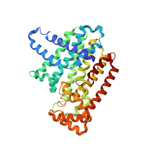
    To understand the structural basis for bisphosphonate therapy of bone diseases, we solved the crystal structures of human farnesyl pyrophosphate synthase (FPPS) in its unliganded state, in complex with the nitrogen-containing bisphosphonate (N-BP) drugs zoledronate, pamidronate, alendronate, and ibandronate, and in the ternary complex with zoledronate and the substrate isopentenyl pyrophosphate (IPP). By revealing three structural snapshots of the enzyme catalytic cycle, each associated with a distinct conformational state, and details about the interactions with N-BPs, these structures provide a novel understanding of the mechanism of FPPS catalysis and inhibition. In particular, the accumulating substrate, IPP, was found to bind to and stabilize the FPPS-N-BP complexes rather than to compete with and displace the N-BP inhibitor. Stabilization of the FPPS-N-BP complex through IPP binding is supported by differential scanning calorimetry analyses of a set of representative N-BPs. Among other factors such as high binding affinity for bone mineral, this particular mode of FPPS inhibition contributes to the exceptional in vivo efficacy of N-BP drugs. Moreover, our data form the basis for structure-guided design of optimized N-BPs with improved pharmacological properties.


  • Organizational Affiliation

    Novartis Institutes for BioMedical Research, Discovery Technologies, 4002 Basel, Switzerland. jeanmichel.rondeau@novartis.com


Macromolecules
Find similar proteins by:  (by identity cutoff)  |  3D Structure
Entity ID: 1
MoleculeChains Sequence LengthOrganismDetailsImage
Farnesyl Diphosphate SynthaseA [auth F]350Homo sapiensMutation(s): 0 
Gene Names: FDPSFPSKIAA1293
EC: 2.5.1.1 (PDB Primary Data), 2.5.1.10 (PDB Primary Data)
UniProt & NIH Common Fund Data Resources
Find proteins for P14324 (Homo sapiens)
Explore P14324 
Go to UniProtKB:  P14324
PHAROS:  P14324
GTEx:  ENSG00000160752 
Entity Groups  
Sequence Clusters30% Identity50% Identity70% Identity90% Identity95% Identity100% Identity
UniProt GroupP14324
Sequence Annotations
Expand
  • Reference Sequence
Small Molecules
Binding Affinity Annotations 
IDSourceBinding Affinity
210 BindingDB:  2F89 Ki: min: 55.9, max: 331.4 (nM) from 3 assay(s)
IC50: min: 200, max: 1932 (nM) from 5 assay(s)
PDBBind:  2F89 Ki: 331 (nM) from 1 assay(s)
Experimental Data & Validation

Experimental Data

  • Method: X-RAY DIFFRACTION
  • Resolution: 2.60 Å
  • R-Value Free: 0.267 
  • R-Value Work: 0.199 
  • R-Value Observed: 0.206 
  • Space Group: P 41 21 2
Unit Cell:
Length ( Å )Angle ( ˚ )
a = 111.569α = 90
b = 111.569β = 90
c = 66.482γ = 90
Software Package:
Software NamePurpose
DENZOdata reduction
SCALEPACKdata scaling
CNXrefinement
CNXphasing

Structure Validation

View Full Validation Report


Ligand Structure Quality Assessment 


Entry History 

Deposition Data

Revision History  (Full details and data files)

  • Version 1.0: 2006-02-28
    Type: Initial release
  • Version 1.1: 2008-05-01
    Changes: Version format compliance
  • Version 1.2: 2011-07-13
    Changes: Derived calculations, Version format compliance
  • Version 1.3: 2017-10-18
    Changes: Refinement description
  • Version 1.4: 2024-02-14
    Changes: Data collection, Database references, Derived calculations