2EFV

Crystal Structure of a Hypothetical Protein(MJ0366) from Methanocaldococcus jannaschii


Experimental Data Snapshot

  • Method: X-RAY DIFFRACTION
  • Resolution: 1.90 Å
  • R-Value Free: 0.231 
  • R-Value Work: 0.219 
  • R-Value Observed: 0.219 

wwPDB Validation   3D Report Full Report


This is version 1.3 of the entry. See complete history


Literature

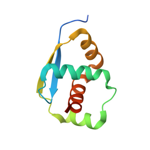
Crystal structure analysis of a hypothetical protein (MJ0366) from Methanocaldococcus jannaschii revealed a novel topological arrangement of the knot fold

Thiruselvam, V.Kumarevel, T.Karthe, P.Kuramitsu, S.Yokoyama, S.Ponnuswamy, M.N.

(2017) Biochem Biophys Res Commun 482: 264-269

  • DOI: https://doi.org/10.1016/j.bbrc.2016.11.052
  • Primary Citation of Related Structures:  
    2EFV

  • PubMed Abstract: 

    The crystal structure of a hypothetical protein MJ0366, derived from Methanocaldococcus jannaschii was solved at 1.9 Å resolution using synchrotron radiation. MJ0366 was crystallized as a monomer and has knot structural arrangement. Intriguingly, the solved structure consists of novel 'KNOT' fold conformation. The 3 1 trefoil knot was observed in the structure. The N-terminal and C-terminal ends did not participate in knot formation.


  • Organizational Affiliation

    Centre of Advanced Study in Crystallography and Biophysics, University of Madras, Guindy Campus, Chennai, 600025, India.


Macromolecules
Find similar proteins by:  (by identity cutoff)  |  3D Structure
Entity ID: 1
MoleculeChains Sequence LengthOrganismDetailsImage
Hypothetical protein MJ036692Methanocaldococcus jannaschii DSM 2661Mutation(s): 0 
Gene Names: mj0366
UniProt
Find proteins for Q57812 (Methanocaldococcus jannaschii (strain ATCC 43067 / DSM 2661 / JAL-1 / JCM 10045 / NBRC 100440))
Explore Q57812 
Go to UniProtKB:  Q57812
Entity Groups  
Sequence Clusters30% Identity50% Identity70% Identity90% Identity95% Identity100% Identity
UniProt GroupQ57812
Sequence Annotations
Expand
  • Reference Sequence
Small Molecules
Ligands 1 Unique
IDChains Name / Formula / InChI Key2D Diagram3D Interactions
PO4
Query on PO4

Download Ideal Coordinates CCD File 
B [auth A]PHOSPHATE ION
O4 P
NBIIXXVUZAFLBC-UHFFFAOYSA-K
Experimental Data & Validation

Experimental Data

  • Method: X-RAY DIFFRACTION
  • Resolution: 1.90 Å
  • R-Value Free: 0.231 
  • R-Value Work: 0.219 
  • R-Value Observed: 0.219 
  • Space Group: P 43 21 2
Unit Cell:
Length ( Å )Angle ( ˚ )
a = 78.846α = 90
b = 78.846β = 90
c = 35.104γ = 90
Software Package:
Software NamePurpose
CNSrefinement
BSSdata collection
HKL-2000data reduction
HKL-2000data scaling
SOLVEphasing

Structure Validation

View Full Validation Report


Entry History 

Deposition Data

Revision History  (Full details and data files)

  • Version 1.0: 2007-08-28
    Type: Initial release
  • Version 1.1: 2011-07-13
    Changes: Version format compliance
  • Version 1.2: 2019-01-16
    Changes: Data collection, Database references
  • Version 1.3: 2024-03-13
    Changes: Data collection, Database references, Derived calculations