2DEM

Crystal structure of Uracil-DNA glycosylase in complex with AP:A containing DNA


Experimental Data Snapshot

  • Method: X-RAY DIFFRACTION
  • Resolution: 1.95 Å
  • R-Value Free: 0.225 
  • R-Value Work: 0.200 

wwPDB Validation   3D Report Full Report


This is version 1.5 of the entry. See complete history


Literature

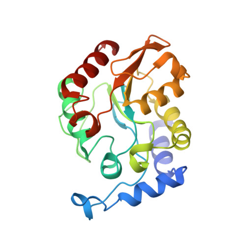
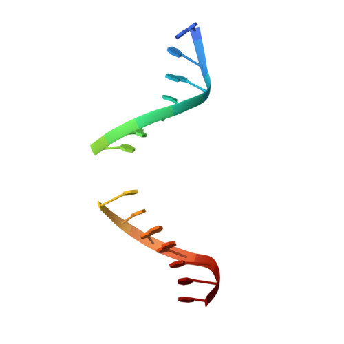
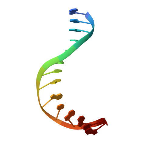
Crystal structure of family 5 uracil-DNA glycosylase bound to DNA.

Kosaka, H.Hoseki, J.Nakagawa, N.Kuramitsu, S.Masui, R.

(2007) J Mol Biol 373: 839-850

  • DOI: https://doi.org/10.1016/j.jmb.2007.08.022
  • Primary Citation of Related Structures:  
    2D3Y, 2DDG, 2DEM

  • PubMed Abstract: 

    Uracil-DNA glycosylase (UDG) removes uracil generated by the deamination of cytosine or misincorporation of deoxyuridine monophosphate. Within the UDG superfamily, a fifth UDG family lacks a polar residue in the active-site motif, which mediates the hydrolysis of the glycosidic bond by activation of a water molecule in UDG families 1-4. We have determined the crystal structure of a novel family 5 UDG from Thermus thermophilus HB8 complexed with DNA containing an abasic site. The active-site structure suggests this enzyme uses both steric force and water activation for its excision reaction. A conserved asparagine residue acts as a ligand to the catalytic water molecule. The structure also implies that another water molecule acts as a barrier during substrate recognition. Based on no significant open-closed conformational change upon binding to DNA, we propose a "slide-in" mechanism for initial damage recognition.


  • Organizational Affiliation

    Department of Biological Sciences, Graduate School of Science, Osaka University, 1-1 Machikaneyama-cho, Toyonaka, Osaka 560-0043, Japan.


Macromolecules

Find similar proteins by:  (by identity cutoff)  |  3D Structure
Entity ID: 3
MoleculeChains Sequence LengthOrganismDetailsImage
uracil-DNA glycosylaseC [auth A]219Thermus thermophilus HB8Mutation(s): 0 
Gene Names: ttUDGB
EC: 3.2.2
UniProt
Find proteins for Q5SJ65 (Thermus thermophilus (strain ATCC 27634 / DSM 579 / HB8))
Explore Q5SJ65 
Go to UniProtKB:  Q5SJ65
Entity Groups  
Sequence Clusters30% Identity50% Identity70% Identity90% Identity95% Identity100% Identity
UniProt GroupQ5SJ65
Sequence Annotations
Expand
  • Reference Sequence

Find similar nucleic acids by:  Sequence   |   3D Structure  

Entity ID: 1
MoleculeChains LengthOrganismImage
5'-D(*AP*TP*GP*TP*TP*GP*CP*(D1P)P*TP*TP*AP*GP*TP*CP*C)-3'A [auth C]15synthetic construct
Sequence Annotations
Expand
  • Reference Sequence

Find similar nucleic acids by:  Sequence   |   3D Structure  

Entity ID: 2
MoleculeChains LengthOrganismImage
5'-D(*GP*GP*AP*CP*TP*AP*AP*AP*GP*CP*AP*AP*CP*A)-3'B [auth D]14synthetic construct
Sequence Annotations
Expand
  • Reference Sequence
Experimental Data & Validation

Experimental Data

  • Method: X-RAY DIFFRACTION
  • Resolution: 1.95 Å
  • R-Value Free: 0.225 
  • R-Value Work: 0.200 
  • Space Group: C 2 2 21
Unit Cell:
Length ( Å )Angle ( ˚ )
a = 68.92α = 90
b = 150.17β = 90
c = 93.41γ = 90
Software Package:
Software NamePurpose
MLPHAREphasing
CNSrefinement

Structure Validation

View Full Validation Report


Entry History 

Deposition Data

Revision History  (Full details and data files)

  • Version 1.0: 2007-04-24
    Type: Initial release
  • Version 1.1: 2008-04-01
    Changes: Version format compliance
  • Version 1.2: 2011-07-13
    Changes: Source and taxonomy, Version format compliance
  • Version 1.3: 2017-10-11
    Changes: Refinement description
  • Version 1.4: 2021-02-17
    Changes: Derived calculations, Source and taxonomy
  • Version 1.5: 2023-10-25
    Changes: Data collection, Database references, Refinement description