2D2M

Structure of an extracellular giant hemoglobin of the gutless beard worm Oligobrachia mashikoi


Experimental Data Snapshot

  • Method: X-RAY DIFFRACTION
  • Resolution: 2.85 Å
  • R-Value Free: 0.277 
  • R-Value Work: 0.236 

wwPDB Validation   3D Report Full Report


This is version 1.2 of the entry. See complete history


Literature

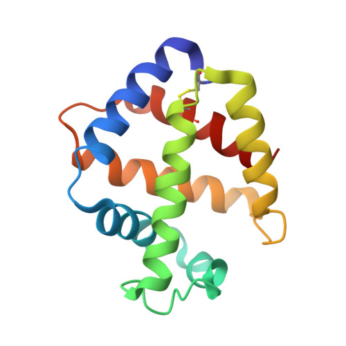
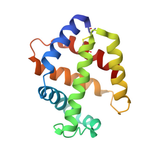
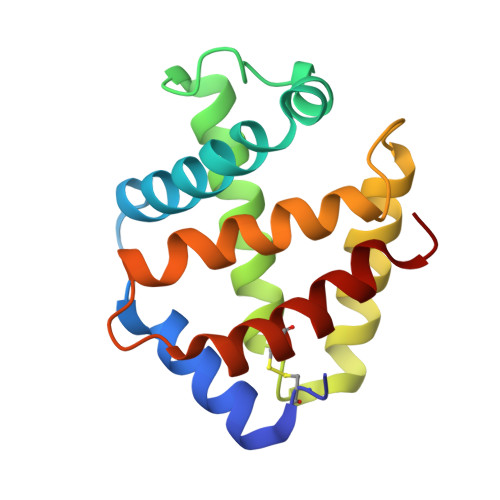
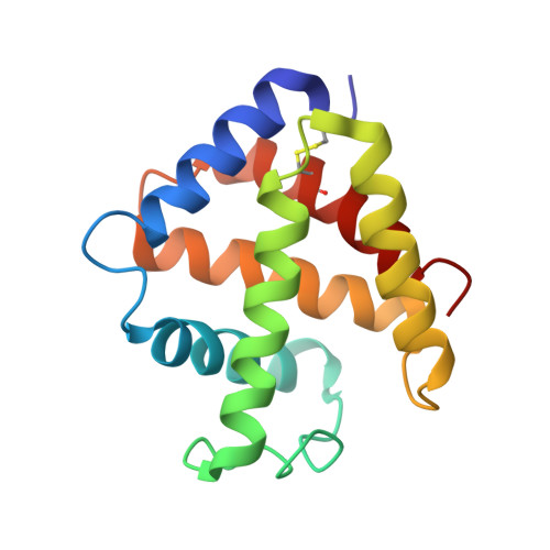
Structure of an extracellular giant hemoglobin of the gutless beard worm Oligobrachia mashikoi.

Numoto, N.Nakagawa, T.Kita, A.Sasayama, Y.Fukumori, Y.Miki, K.

(2005) Proc Natl Acad Sci U S A 102: 14521-14526

  • DOI: https://doi.org/10.1073/pnas.0501541102
  • Primary Citation of Related Structures:  
    2D2M, 2D2N

  • PubMed Abstract: 

    Mouthless and gutless marine animals, pogonophorans and vestimentiferans, obtain their nutrition solely from their symbiotic chemoautotrophic sulfur-oxidizing bacteria. These animals have sulfide-binding 400-kDa and/or 3,500-kDa Hb, which transports oxygen and sulfide simultaneously. The symbiotic bacteria are supplied with sulfide by Hb of the host animal and use it to provide carbon compounds. Here, we report the crystal structure of a 400-kDa Hb from pogonophoran Oligobrachia mashikoi at 2.85-A resolution. The structure is hollow-spherical, composed of a total of 24 globins as a dimer of dodecamer. This dodecameric assemblage would be a fundamental structural unit of both 400-kDa and 3,500-kDa Hbs. The structure of the mercury derivative used for phasing provides insights into the sulfide-binding mechanism. The mercury compounds bound to all free Cys residues that have been expected as sulfide-binding sites. Some of the free Cys residues are surrounded by Phe aromatic rings, and mercury atoms come into contact with these residues in the derivative structure. It is strongly suggested that sulfur atoms bound to these sites could be stabilized by aromatic-electrostatic interactions by the surrounding Phe residues.


  • Organizational Affiliation

    Department of Chemistry, Graduate School of Science, Kyoto University, Sakyo-ku, Kyoto 606-8502, Japan.


Macromolecules
Find similar proteins by:  (by identity cutoff)  |  3D Structure
Entity ID: 1
MoleculeChains Sequence LengthOrganismDetailsImage
Giant hemoglobin, A1(b) globin chain140Oligobrachia mashikoiMutation(s): 0 
UniProt
Find proteins for Q7M419 (Oligobrachia mashikoi)
Explore Q7M419 
Go to UniProtKB:  Q7M419
Entity Groups  
Sequence Clusters30% Identity50% Identity70% Identity90% Identity95% Identity100% Identity
UniProt GroupQ7M419
Sequence Annotations
Expand
  • Reference Sequence
Find similar proteins by:  (by identity cutoff)  |  3D Structure
Entity ID: 2
MoleculeChains Sequence LengthOrganismDetailsImage
Giant hemoglobin, A2(a5) globin chain142Oligobrachia mashikoiMutation(s): 0 
UniProt
Find proteins for Q7M413 (Oligobrachia mashikoi)
Explore Q7M413 
Go to UniProtKB:  Q7M413
Entity Groups  
Sequence Clusters30% Identity50% Identity70% Identity90% Identity95% Identity100% Identity
UniProt GroupQ7M413
Sequence Annotations
Expand
  • Reference Sequence
Find similar proteins by:  (by identity cutoff)  |  3D Structure
Entity ID: 3
MoleculeChains Sequence LengthOrganismDetailsImage
Giant hemoglobin, B2(c) globin chain147Oligobrachia mashikoiMutation(s): 0 
UniProt
Find proteins for Q7M418 (Oligobrachia mashikoi)
Explore Q7M418 
Go to UniProtKB:  Q7M418
Entity Groups  
Sequence Clusters30% Identity50% Identity70% Identity90% Identity95% Identity100% Identity
UniProt GroupQ7M418
Sequence Annotations
Expand
  • Reference Sequence
Find similar proteins by:  (by identity cutoff)  |  3D Structure
Entity ID: 4
MoleculeChains Sequence LengthOrganismDetailsImage
Giant hemoglobin, B1(d) globin chain145Oligobrachia mashikoiMutation(s): 0 
UniProt
Find proteins for Q5KSB7 (Oligobrachia mashikoi)
Explore Q5KSB7 
Go to UniProtKB:  Q5KSB7
Entity Groups  
Sequence Clusters30% Identity50% Identity70% Identity90% Identity95% Identity100% Identity
UniProt GroupQ5KSB7
Sequence Annotations
Expand
  • Reference Sequence
Experimental Data & Validation

Experimental Data

  • Method: X-RAY DIFFRACTION
  • Resolution: 2.85 Å
  • R-Value Free: 0.277 
  • R-Value Work: 0.236 
  • Space Group: H 3 2
Unit Cell:
Length ( Å )Angle ( ˚ )
a = 111.498α = 90
b = 111.498β = 90
c = 276.835γ = 120
Software Package:
Software NamePurpose
CNSrefinement
HKL-2000data reduction
SCALEPACKdata scaling
SOLVEphasing

Structure Validation

View Full Validation Report


Entry History 

Deposition Data

Revision History  (Full details and data files)

  • Version 1.0: 2005-10-25
    Type: Initial release
  • Version 1.1: 2008-04-30
    Changes: Version format compliance
  • Version 1.2: 2011-07-13
    Changes: Version format compliance