2CYF

The Crystal Structure of Canavalia Maritima Lectin (ConM) in Complex with Trehalose and Maltose


Experimental Data Snapshot

  • Method: X-RAY DIFFRACTION
  • Resolution: 1.80 Å
  • R-Value Free: 0.238 
  • R-Value Work: 0.186 
  • R-Value Observed: 0.194 

wwPDB Validation   3D Report Full Report


This is version 2.1 of the entry. See complete history


Literature

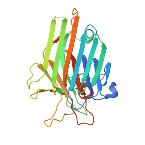
Crystal structure of a lectin from Canavalia maritima (ConM) in complex with trehalose and maltose reveals relevant mutation in ConA-like lectins

Delatorre, P.Rocha, B.A.M.Gadelha, C.A.A.Santi-Gadelha, T.Cajazeiras, J.B.Souza, E.P.Nascimento, K.S.Freire, V.N.Sampaio, A.H.Azevedo Jr., W.F.Cavada, B.S.

(2006) J Struct Biol 154: 280-286

  • DOI: https://doi.org/10.1016/j.jsb.2006.03.011
  • Primary Citation of Related Structures:  
    2CY6, 2CYF

  • PubMed Abstract: 

    The crystal structure of Canavalia maritima lectin (ConM) complexed with trehalose and maltose revealed relevant point mutations in ConA-like lectins. ConM with the disaccharides and other ConA-like lectins complexed with carbohydrates demonstrated significant differences in the position of H-bonds. The main difference in the ConM structure is the replacement of Pro202 by Ser202, a residue that promotes the approximation of Tyr12 to the carbohydrate-binding site. The O-6' of the second glucose ring in maltose interacts with Tyr12, while in trehalose the interaction is established by the O-2' and Tyr12, explaining the higher affinity of ConM for disaccharides compared to monosaccharides.


  • Organizational Affiliation

    Departamento de Bioquímica e Biologia Molecular--Universidade Federal do Ceará, Brazil.


Macromolecules
Find similar proteins by:  (by identity cutoff)  |  3D Structure
Entity ID: 1
MoleculeChains Sequence LengthOrganismDetailsImage
LectinA,
B [auth C]
237Canavalia roseaMutation(s): 0 
UniProt
Find proteins for P55915 (Canavalia brasiliensis)
Explore P55915 
Go to UniProtKB:  P55915
Entity Groups  
Sequence Clusters30% Identity50% Identity70% Identity90% Identity95% Identity100% Identity
UniProt GroupP55915
Sequence Annotations
Expand
  • Reference Sequence
Oligosaccharides

Help

Entity ID: 2
MoleculeChains Length2D Diagram Glycosylation3D Interactions
alpha-D-glucopyranose-(1-4)-alpha-D-glucopyranoseC [auth B],
D
2N/A
Glycosylation Resources
GlyTouCan:  G07411ON
GlyCosmos:  G07411ON
Biologically Interesting Molecules (External Reference) 1 Unique
Experimental Data & Validation

Experimental Data

  • Method: X-RAY DIFFRACTION
  • Resolution: 1.80 Å
  • R-Value Free: 0.238 
  • R-Value Work: 0.186 
  • R-Value Observed: 0.194 
  • Space Group: P 21 21 2
Unit Cell:
Length ( Å )Angle ( ˚ )
a = 67.033α = 90
b = 97.332β = 90
c = 71.086γ = 90
Software Package:
Software NamePurpose
REFMACrefinement
MAR345data collection
CCP4data scaling
AMoREphasing

Structure Validation

View Full Validation Report


Entry History 

Deposition Data

Revision History  (Full details and data files)

  • Version 1.0: 2006-06-13
    Type: Initial release
  • Version 1.1: 2008-04-30
    Changes: Version format compliance
  • Version 1.2: 2011-07-13
    Changes: Version format compliance
  • Version 1.3: 2017-10-11
    Changes: Advisory, Refinement description
  • Version 2.0: 2020-07-29
    Type: Remediation
    Reason: Carbohydrate remediation
    Changes: Advisory, Atomic model, Data collection, Derived calculations, Non-polymer description, Structure summary
  • Version 2.1: 2024-03-13
    Changes: Data collection, Database references, Structure summary