2CFH

Structure of the Bet3-TPC6B core of TRAPP


Experimental Data Snapshot

  • Method: X-RAY DIFFRACTION
  • Resolution: 2.30 Å
  • R-Value Free: 0.269 
  • R-Value Work: 0.221 
  • R-Value Observed: 0.223 

wwPDB Validation 

Currently 2CFH does not have a validation slider image.


This is version 1.3 of the entry. See complete history


Literature

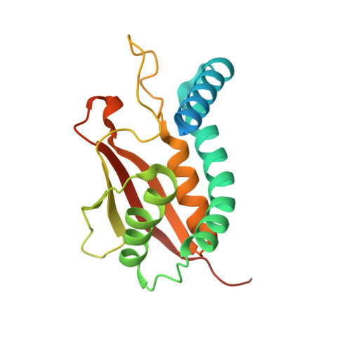
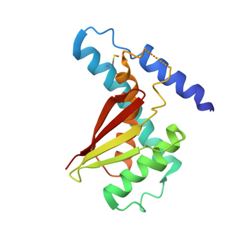
Structure of the Bet3-Tpc6B Core of Trapp: Two Tpc6 Paralogs Form Trimeric Complexes with Bet3 and Mum2.

Kummel, D.Muller, J.J.Roske, Y.Henke, N.Heinemann, U.

(2006) J Mol Biol 361: 22

  • DOI: https://doi.org/10.1016/j.jmb.2006.06.012
  • Primary Citation of Related Structures:  
    2CFH

  • PubMed Abstract: 

    The transport protein particle (TRAPP) complexes are involved in the tethering process at different trafficking steps of vesicle transport. We here present the crystal structure of a human Bet3-Tpc6B heterodimer, which represents a core sub-complex in the assembly of TRAPP. We describe a conserved patch of Tpc6 with uncharged pockets, forming a putative interaction interface for an anchoring moiety at the Golgi. The structural and functional comparison of the two paralogs Tpc6A and Tpc6B, only found in some organisms, indicates redundancy and added complexity of TRAPP architecture and function. Both iso-complexes, Bet3-Tpc6A and Bet3-Tpc6B, are able to recruit Mum2, a further TRAPP subunit, and we identify the alpha1-alpha2 loop regions as a binding site for Mum2. Our study reveals similar stability of the iso-complexes and similar expression patterns of the tpc6 variants in different mouse organs. These findings raise the possibility that the Tpc6 paralogs might contribute to the formation of two distinct TRAPP complexes that differ in function.


  • Organizational Affiliation

    Max-Delbrück Center for Molecular Medicine, Berlin, Germany.


Macromolecules
Find similar proteins by:  (by identity cutoff)  |  3D Structure
Entity ID: 1
MoleculeChains Sequence LengthOrganismDetailsImage
TRAFFICKING PROTEIN PARTICLE COMPLEX SUBUNIT 3
A, B
194Homo sapiensMutation(s): 0 
UniProt & NIH Common Fund Data Resources
Find proteins for O43617 (Homo sapiens)
Explore O43617 
Go to UniProtKB:  O43617
PHAROS:  O43617
GTEx:  ENSG00000054116 
Entity Groups  
Sequence Clusters30% Identity50% Identity70% Identity90% Identity95% Identity100% Identity
UniProt GroupO43617
Sequence Annotations
Expand
  • Reference Sequence
Find similar proteins by:  (by identity cutoff)  |  3D Structure
Entity ID: 2
MoleculeChains Sequence LengthOrganismDetailsImage
TRAFFICKING PROTEIN PARTICLE COMPLEX SUBUNIT 6B
C, D
158Homo sapiensMutation(s): 0 
UniProt & NIH Common Fund Data Resources
Find proteins for Q86SZ2 (Homo sapiens)
Explore Q86SZ2 
Go to UniProtKB:  Q86SZ2
PHAROS:  Q86SZ2
GTEx:  ENSG00000182400 
Entity Groups  
Sequence Clusters30% Identity50% Identity70% Identity90% Identity95% Identity100% Identity
UniProt GroupQ86SZ2
Sequence Annotations
Expand
  • Reference Sequence
Small Molecules
Modified Residues  1 Unique
IDChains TypeFormula2D DiagramParent
CSO
Query on CSO
C, D
L-PEPTIDE LINKINGC3 H7 N O3 SCYS
Experimental Data & Validation

Experimental Data

  • Method: X-RAY DIFFRACTION
  • Resolution: 2.30 Å
  • R-Value Free: 0.269 
  • R-Value Work: 0.221 
  • R-Value Observed: 0.223 
  • Space Group: C 1 2 1
Unit Cell:
Length ( Å )Angle ( ˚ )
a = 69.45α = 90
b = 69.59β = 91.55
c = 144.006γ = 90
Software Package:
Software NamePurpose
REFMACrefinement
XDSdata reduction
XSCALEdata scaling
MOLREPphasing

Structure Validation

Currently 2CFH does not have a validation slider image.


Entry History 

Deposition Data

Revision History  (Full details and data files)

  • Version 1.0: 2006-07-12
    Type: Initial release
  • Version 1.1: 2011-07-13
    Changes: Advisory, Version format compliance
  • Version 1.2: 2011-10-26
    Changes: Atomic model, Derived calculations, Non-polymer description, Other, Refinement description, Structure summary
  • Version 1.3: 2023-12-13
    Changes: Data collection, Database references, Derived calculations, Other, Refinement description