Conservation of Structure and Mechanism in Primary and Secondary Transporters Exemplified by Siap, a Sialic Acid Binding Virulence Factor from Haemophilus Influenzae
Muller, A., Severi, E., Mulligan, C., Watts, A.G., Kelly, D.J., Wilson, K.S., Wilkinson, A.J., Thomas, G.H.(2006) J Biol Chem 281: 22212
- PubMed: 16702222
- DOI: https://doi.org/10.1074/jbc.M603463200
- Primary Citation of Related Structures:
2CEX, 2CEY - PubMed Abstract:
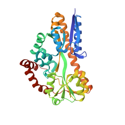
Extracytoplasmic solute receptors (ESRs) are important components of solute uptake systems in bacteria, having been studied extensively as parts of ATP binding cassette transporters. Herein we report the first crystal structure of an ESR protein from a functionally characterized electrochemical ion gradient dependent secondary transporter. This protein, SiaP, forms part of a tripartite ATP-independent periplasmic transporter specific for sialic acid in Haemophilus influenzae. Surprisingly, the structure reveals an overall topology similar to ATP binding cassette ESR proteins, which is not apparent from the sequence, demonstrating that primary and secondary transporters can share a common structural component. The structure of SiaP in the presence of the sialic acid analogue 2,3-didehydro-2-deoxy-N-acetylneuraminic acid reveals the ligand bound in a deep cavity with its carboxylate group forming a salt bridge with a highly conserved Arg residue. Sialic acid binding, which obeys simple bimolecular association kinetics as determined by stopped-flow fluorescence spectroscopy, is accompanied by domain closure about a hinge region and the kinking of an alpha-helix hinge component. The structure provides insight into the evolution, mechanism, and substrate specificity of ESR-dependent secondary transporters that are widespread in prokaryotes.
Organizational Affiliation:
Structural Biology Laboratory, Department of Chemistry, University of York, York YO10 5YW, United Kingdom.