2CBQ

Crystal structure of the neocarzinostatin 1Tes15 mutant bound to testosterone hemisuccinate.


Experimental Data Snapshot

  • Method: X-RAY DIFFRACTION
  • Resolution: 2.60 Å
  • R-Value Free: 0.257 
  • R-Value Work: 0.207 
  • R-Value Observed: 0.207 

wwPDB Validation   3D Report Full Report


This is version 1.3 of the entry. See complete history


Literature

Structures of in Vitro Evolved Binding Sites on Neocarzinostatin Scaffold Reveal Unanticipated Evolutionary Pathways.

Drevelle, A.Graille, M.Heyd, B.Sorel, I.Ulryck, N.Pecorari, F.Desmadril, M.Van Tilbeurgh, H.Minard, P.

(2006) J Mol Biol 358: 455

  • DOI: https://doi.org/10.1016/j.jmb.2006.02.002
  • Primary Citation of Related Structures:  
    2CBM, 2CBO, 2CBQ, 2CBT

  • PubMed Abstract: 

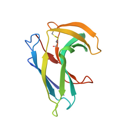
    We have recently applied in vitro evolution methods to create in Neocarzinostatin a new binding site for a target molecule unrelated to its natural ligand. The main objective of this work was to solve the structure of some of the selected binders in complex with the target molecule: testosterone. Three proteins (1a.15, 3.24 and 4.1) were chosen as representative members of sequence families that came out of the selection process within different randomization schemes. In order to evaluate ligand-induced conformational adaptation, we also determined the structure of one of the proteins (3.24) in the free and complexed forms. Surprisingly, all these mutants bind not one but two molecules of testosterone in two very different ways. The 3.24 structure revealed that the protein spontaneously evolved in the system to bind two ligand molecules in one single binding crevice. These two binding sites are formed by substituted as well as by non-variable side-chains. The comparison with the free structure shows that only limited structural changes are observed upon ligand binding. The X-ray structures of the complex formed by 1a.15 and 4.1 Neocarzinostatin mutants revealed that the two variants form very similar dimers. These dimers were observed neither for the uncomplexed variants nor for wild-type Neocarzinostatin but were shown here to be induced by ligand binding. Comparison of the three complexed forms clearly suggests that these unanticipated structural responses resulted from the molecular arrangement used for the selection experiments.


  • Organizational Affiliation

    Equipe de Modélisation et Ingénierie des Protéines, IBBMC, CNRS, UMR8619, Université Paris XI, Bât. 430, 91405 Orsay, France.


Macromolecules
Find similar proteins by:  (by identity cutoff)  |  3D Structure
Entity ID: 1
MoleculeChains Sequence LengthOrganismDetailsImage
NEOCARZINOSTATIN
A, B, C, D, E
A, B, C, D, E, F
114Streptomyces carzinostaticusMutation(s): 7 
UniProt
Find proteins for P0A3R9 (Streptomyces carzinostaticus)
Explore P0A3R9 
Go to UniProtKB:  P0A3R9
Entity Groups  
Sequence Clusters30% Identity50% Identity70% Identity90% Identity95% Identity100% Identity
UniProt GroupP0A3R9
Sequence Annotations
Expand
  • Reference Sequence
Small Molecules
Ligands 2 Unique
IDChains Name / Formula / InChI Key2D Diagram3D Interactions
TH2
Query on TH2

Download Ideal Coordinates CCD File 
H [auth A]
I [auth A]
K [auth B]
L [auth B]
N [auth C]
H [auth A],
I [auth A],
K [auth B],
L [auth B],
N [auth C],
O [auth D],
Q [auth E],
S [auth F]
TESTOSTERONE HEMISUCCINATE
C23 H32 O5
CJQNBXFUHQZFOE-VYAQIDIUSA-N
SO4
Query on SO4

Download Ideal Coordinates CCD File 
G [auth A],
J [auth B],
M [auth C],
P [auth E],
R [auth F]
SULFATE ION
O4 S
QAOWNCQODCNURD-UHFFFAOYSA-L
Experimental Data & Validation

Experimental Data

  • Method: X-RAY DIFFRACTION
  • Resolution: 2.60 Å
  • R-Value Free: 0.257 
  • R-Value Work: 0.207 
  • R-Value Observed: 0.207 
  • Space Group: P 21 21 21
Unit Cell:
Length ( Å )Angle ( ˚ )
a = 52.976α = 90
b = 97.827β = 90
c = 129.268γ = 90
Software Package:
Software NamePurpose
CNSrefinement
DENZOdata reduction
SCALEPACKdata scaling
AMoREphasing

Structure Validation

View Full Validation Report


Entry History 

Deposition Data

Revision History  (Full details and data files)

  • Version 1.0: 2006-03-22
    Type: Initial release
  • Version 1.1: 2011-05-08
    Changes: Version format compliance
  • Version 1.2: 2011-07-13
    Changes: Version format compliance
  • Version 1.3: 2023-12-13
    Changes: Data collection, Database references, Other, Refinement description