The Crystal Structure of the Zinc Phosphodiesterase from Escherichia Coli Provides Insight Into Function and Cooperativity of Trnase Z-Family Proteins.
Kostelecky, B., Pohl, E., Vogel, A., Schilling, O., Meyer-Klaucke, W.(2006) J Bacteriol 188: 1607
- PubMed: 16452444
- DOI: https://doi.org/10.1128/JB.188.4.1607-1614.2006
- Primary Citation of Related Structures:
2CBN - PubMed Abstract:
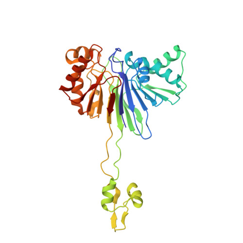
The elaC gene product from Escherichia coli, ZiPD, is a 3' tRNA-processing endonuclease belonging to the tRNase Z family of enzymes that have been identified in a wide variety of organisms. In contrast to the elaC homologue from Bacillus subtilis, E. coli elaC is not essential for viability, and although both enzymes process only precursor tRNA (pre-tRNA) lacking a CCA triplet at the 3' end in vitro, the physiological role of ZiPD remains enigmatic because all pre-tRNA species in E. coli are transcribed with the CCA triplet. We present the first crystal structure of ZiPD determined by multiple anomalous diffraction at a resolution of 2.9 A. This structure shares many features with the tRNase Z enzymes from B. subtilis and Thermotoga maritima, but there are distinct differences in metal binding and overall domain organization. Unlike the previously described homologous structures, ZiPD dimers display crystallographic symmetry and fully loaded metal sites. The ZiPD exosite is similar to that of the B. subtilis enzyme structurally, but its position with respect to the protein core differs substantially, illustrating its ability to act as a clamp in binding tRNA. Furthermore, the ZiPD crystal structure presented here provides insight into the enzyme's cooperativity and assists the ongoing attempt to elucidate the physiological function of this protein.
Organizational Affiliation:
EMBL Hamburg Outstation c/o DESY, Notkestrasse 85, D-22603 Hamburg, Germany.