Determination of the three-dimensional solution structure of the C-terminal domain of cellobiohydrolase I from Trichoderma reesei. A study using nuclear magnetic resonance and hybrid distance geometry-dynamical simulated annealing.
Kraulis, J., Clore, G.M., Nilges, M., Jones, T.A., Pettersson, G., Knowles, J., Gronenborn, A.M.(1989) Biochemistry 28: 7241-7257
- PubMed: 2554967
- DOI: https://doi.org/10.1021/bi00444a016
- Primary Citation of Related Structures:
1CBH, 2CBH - PubMed Abstract:
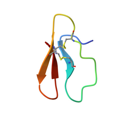
The solution structure of a synthetic 36-residue polypeptide comprising the C-terminal cellulose binding domain of cellobiohydrolase I (CT-CBH I) from Trichoderma reesei was investigated by nuclear magnetic resonance (NMR) spectroscopy. The 1H NMR spectrum was completely assigned in a sequential manner by two-dimensional NMR techniques. A large number of stereospecific assignments for beta-methylene protons, as well as ranges for the phi, psi, and chi 1 torsion angles, were obtained on the basis of sequential and intraresidue nuclear Overhauser enhancement (NOE) and coupling constant data in combination with a conformational data base search. The structure calculations were carried out in an iterative manner by using the hybrid distance geometry-dynamical simulated annealing method. This involved computing a series of initial structures from a subset of the experimental data in order to resolve ambiguities in the assignments of some NOE cross-peaks arising from chemical shift degeneracy. Additionally, this permitted us to extend the stereospecific assignments to the alpha-methylene protons of glycine using information on phi torsion angles derived from the initial structure calculations. The final experimental data set consisted of 554 interproton distance restraints, 24 restraints for 12 hydrogen bonds, and 33 phi, 24 psi, and 25 chi 1 torsion angle restraints. CT-CBH I has two disulfide bridges whose pairing was previously unknown. Analysis of structures calculated with all three possible combinations of disulfide bonds, as well as without disulfide bonds, indicated that the correct disulfide bridge pairing was 8-25 and 19-35. Forty-one structures were computed with the 8-25 and 19-35 disulfide bridges, and the average atomic rms difference between the individual structures and the mean structure obtained by averaging their coordinates was 0.33 +/- 0.04 A for the backbone atoms and 0.52 +/- 0.06 A for all atoms. The protein has a wedgelike shape with an amphiphilic character, one face being predominantly hydrophilic and the other mainly hydrophobic. The principal element of secondary structure is made up of an irregular triple-stranded antiparallel beta-sheet composed of residues 5-9 (beta 1), 24-28 (beta 2), and 33-36 (beta 3) in which strand beta 3 is hydrogen bonded to the other two strands.(ABSTRACT TRUNCATED AT 400 WORDS)
Organizational Affiliation:
Laboratory of Chemical Physics, National Institute of Diabetes and Digestive and Kidney Diseases, Bethesda, Maryland 20892.