


Crystal Structure of Ferrochelatase Hemh-1 from Bacillus Anthracis, Str. Ames
Muller, A., Lebedev, A.A., Moroz, O.V., Blagova, E.V., Levdikov, V.M., Fogg, M.J., Brannigan, J.A., Wilkinson, A.J., Wilson, K.S.To be published.
Experimental Data Snapshot
wwPDB Validation 3D Report Full Report

Entity ID: 1 | |||||
|---|---|---|---|---|---|
| Molecule | Chains | Sequence Length | Organism | Details | Image |
| FERROCHELATASE 1 | 311 | Bacillus anthracis str. Ames | Mutation(s): 0 EC: 4.99.1.1 | 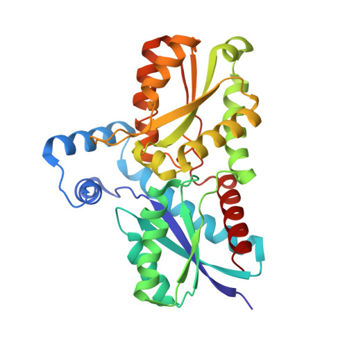 | |
UniProt | |||||
Find proteins for Q81U22 (Bacillus anthracis) Explore Q81U22 Go to UniProtKB: Q81U22 | |||||
Entity Groups | |||||
| Sequence Clusters | 30% Identity50% Identity70% Identity90% Identity95% Identity100% Identity | ||||
| UniProt Group | Q81U22 | ||||
Sequence AnnotationsExpand | |||||
| |||||
| Length ( Å ) | Angle ( ˚ ) |
|---|---|
| a = 49.931 | α = 90 |
| b = 109.927 | β = 90.12 |
| c = 59.404 | γ = 90 |
| Software Name | Purpose |
|---|---|
| REFMAC | refinement |
| MOSFLM | data reduction |
| SCALA | data scaling |
| MOLREP | phasing |
RCSB PDB (citation) is hosted by
RCSB PDB is a member of the

