2C5Q

Crystal structure of yeast YER010Cp


Experimental Data Snapshot

  • Method: X-RAY DIFFRACTION
  • Resolution: 1.70 Å
  • R-Value Free: 0.183 
  • R-Value Work: 0.155 
  • R-Value Observed: 0.156 

wwPDB Validation   3D Report Full Report


This is version 1.2 of the entry. See complete history


Literature

Crystal Structure of Yeast Yer010Cp, a Knotable Member of the Rraa Protein Family.

Leulliot, N.Quevillon-Cheruel, S.Graille, M.Schiltz, M.Blondeau, K.Janin, J.Van Tilbeurgh, H.

(2005) Protein Sci 14: 2751

  • DOI: https://doi.org/10.1110/ps.051684005
  • Primary Citation of Related Structures:  
    2C5Q

  • PubMed Abstract: 

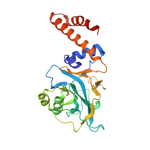
    We present here the structure of Yer010c protein of unknown function, solved by Multiple Anomalous Diffraction and revealing a common fold and oligomerization state with proteins of the regulator of ribonuclease activity A (RraA) family. In Escherichia coli, RraA has been shown to regulate the activity of ribonuclease E by direct interaction. The absence of ribonuclease E in yeast suggests a different function for this family member in this organism. Yer010cp has a few supplementary secondary structure elements and a deep pseudo-knot at the heart of the protein core. A tunnel at the interface between two monomers, lined with conserved charged residues, has unassigned residual electron density and may constitute an active site for a yet unknown activity.


  • Organizational Affiliation

    Institut de Biochimie et de Biophysique Moléculaire et Cellulaire (CNRS-UMR 8619), Université Paris-Sud, Bâtiment 430, 91405 Orsay, France. Nicolas.leulliot@ibbmc.u-psud.fr


Macromolecules
Find similar proteins by:  (by identity cutoff)  |  3D Structure
Entity ID: 1
MoleculeChains Sequence LengthOrganismDetailsImage
RRAA-LIKE PROTEIN YER010C
A, B, C, D, E
A, B, C, D, E, F
240Saccharomyces cerevisiaeMutation(s): 0 
UniProt
Find proteins for P40011 (Saccharomyces cerevisiae (strain ATCC 204508 / S288c))
Explore P40011 
Go to UniProtKB:  P40011
Entity Groups  
Sequence Clusters30% Identity50% Identity70% Identity90% Identity95% Identity100% Identity
UniProt GroupP40011
Sequence Annotations
Expand
  • Reference Sequence
Small Molecules
Ligands 1 Unique
IDChains Name / Formula / InChI Key2D Diagram3D Interactions
EDO
Query on EDO

Download Ideal Coordinates CCD File 
G [auth A]
H [auth B]
I [auth B]
J [auth C]
K [auth D]
G [auth A],
H [auth B],
I [auth B],
J [auth C],
K [auth D],
L [auth D],
M [auth D],
N [auth D],
O [auth F],
P [auth F],
Q [auth F],
R [auth F],
S [auth F]
1,2-ETHANEDIOL
C2 H6 O2
LYCAIKOWRPUZTN-UHFFFAOYSA-N
Modified Residues  1 Unique
IDChains TypeFormula2D DiagramParent
CME
Query on CME
A, B, C, D, E
A, B, C, D, E, F
L-PEPTIDE LINKINGC5 H11 N O3 S2CYS
Experimental Data & Validation

Experimental Data

  • Method: X-RAY DIFFRACTION
  • Resolution: 1.70 Å
  • R-Value Free: 0.183 
  • R-Value Work: 0.155 
  • R-Value Observed: 0.156 
  • Space Group: P 21 21 2
Unit Cell:
Length ( Å )Angle ( ˚ )
a = 128.193α = 90
b = 255.073β = 90
c = 48.173γ = 90
Software Package:
Software NamePurpose
MOSFLMdata reduction
SCALAdata scaling
SHELXDphasing
SHARPphasing
REFMACrefinement

Structure Validation

View Full Validation Report


Entry History 

Deposition Data

Revision History  (Full details and data files)

  • Version 1.0: 2005-11-14
    Type: Initial release
  • Version 1.1: 2011-05-08
    Changes: Version format compliance
  • Version 1.2: 2011-07-13
    Changes: Version format compliance