2BOS

A MUTANT SHIGA-LIKE TOXIN IIE BOUND TO ITS RECEPTOR


Experimental Data Snapshot

  • Method: X-RAY DIFFRACTION
  • Resolution: 2.00 Å
  • R-Value Free: 0.194 
  • R-Value Work: 0.155 
  • R-Value Observed: 0.155 

wwPDB Validation   3D Report Full Report


This is version 2.2 of the entry. See complete history


Literature

A mutant Shiga-like toxin IIe bound to its receptor Gb(3): structure of a group II Shiga-like toxin with altered binding specificity.

Ling, H.Pannu, N.S.Boodhoo, A.Armstrong, G.D.Clark, C.G.Brunton, J.L.Read, R.J.

(2000) Structure 8: 253-264

  • DOI: https://doi.org/10.1016/s0969-2126(00)00103-9
  • Primary Citation of Related Structures:  
    1QOH, 2BOS

  • PubMed Abstract: 

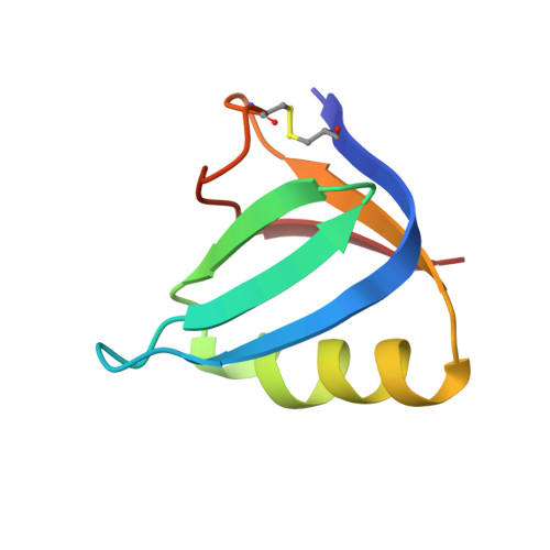
    Shiga-like toxins (SLTs) are produced by the pathogenic strains of Escherichia coli that cause hemorrhagic colitis and hemolytic uremic syndrome. These diseases in humans are generally associated with group II family members (SLT-II and SLT-IIc), whereas SLT-IIe (pig edema toxin) is central to edema disease of swine. The pentameric B-subunit component of the majority of family members binds to the cell-surface glycolipid globotriaosyl ceramide (Gb(3)), but globotetraosyl ceramide (Gb(4)) is the preferred receptor for SLT-IIe. A double-mutant of the SLT-IIe B subunit that reverses two sequence differences from SLT-II (GT3; Gln65-->Glu, Lys67-->Gln, SLT-I numbering) has been shown to bind more strongly to Gb(3) than to Gb(4).


  • Organizational Affiliation

    Department of Biochemistry, University of Alberta, Edmonton, T6G 2H7, Canada.


Macromolecules
Find similar proteins by:  (by identity cutoff)  |  3D Structure
Entity ID: 1
MoleculeChains Sequence LengthOrganismDetailsImage
PROTEIN (SHIGA-LIKE TOXIN IIE B SUBUNIT)
A, B, C, D, E
68Escherichia coliMutation(s): 2 
UniProt
Find proteins for Q47644 (Escherichia coli)
Explore Q47644 
Go to UniProtKB:  Q47644
Entity Groups  
Sequence Clusters30% Identity50% Identity70% Identity90% Identity95% Identity100% Identity
UniProt GroupQ47644
Sequence Annotations
Expand
  • Reference Sequence
Oligosaccharides

Help

Entity ID: 2
MoleculeChains Length2D Diagram Glycosylation3D Interactions
alpha-D-galactopyranose-(1-4)-beta-D-galactopyranose-(1-4)-alpha-D-glucopyranose
F, G, H, J, K
3N/A
Glycosylation Resources
GlyTouCan:  G79094AA
GlyCosmos:  G79094AA
GlyGen:  G79094AA
Entity ID: 3
MoleculeChains Length2D Diagram Glycosylation3D Interactions
alpha-D-galactopyranose-(1-4)-beta-D-galactopyranose
I, L
2N/A
Glycosylation Resources
GlyTouCan:  G67988RO
GlyCosmos:  G67988RO
GlyGen:  G67988RO
Experimental Data & Validation

Experimental Data

  • Method: X-RAY DIFFRACTION
  • Resolution: 2.00 Å
  • R-Value Free: 0.194 
  • R-Value Work: 0.155 
  • R-Value Observed: 0.155 
  • Space Group: P 21 21 21
Unit Cell:
Length ( Å )Angle ( ˚ )
a = 62.077α = 90
b = 78.618β = 90
c = 78.468γ = 90
Software Package:
Software NamePurpose
X-GENdata scaling
X-GENdata reduction
AMoREphasing
BRUTEmodel building
X-PLORrefinement
BRUTEphasing

Structure Validation

View Full Validation Report


Entry History 

Deposition Data

Revision History  (Full details and data files)

  • Version 1.0: 1999-10-20
    Type: Initial release
  • Version 1.1: 2008-04-27
    Changes: Version format compliance
  • Version 1.2: 2011-07-13
    Changes: Version format compliance
  • Version 1.3: 2017-10-04
    Changes: Refinement description
  • Version 2.0: 2020-07-29
    Type: Remediation
    Reason: Carbohydrate remediation
    Changes: Advisory, Atomic model, Data collection, Derived calculations, Structure summary
  • Version 2.1: 2021-11-03
    Changes: Database references, Structure summary
  • Version 2.2: 2023-08-23
    Changes: Data collection, Refinement description