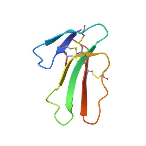
Glycosphingolipid-Facilitated Membrane Insertion and Internalization of Cobra Cardiotoxin: The Sulfatide/Cardiotoxin Complex Structure in a Membrane-Like Environment Suggests a Lipid-Dependent Cell-Penetrating Mechanism for Membrane Binding Polypeptides.
Wang, C.-H., Liu, J.-H., Lee, S.-C., Hsiao, C.-D., Wu, W.-G.(2006) J Biol Chem 281: 656
- PubMed: 16263708
- DOI: https://doi.org/10.1074/jbc.M507880200
- Primary Citation of Related Structures:
2BHI - PubMed Abstract:
Cobra cardiotoxins, a family of basic polypeptides having lipid- and heparin-binding capacities similar to the cell-penetrating peptides, induce severe tissue necrosis and systolic heart arrest in snakebite victims. Whereas cardiotoxins are specifically retained on the cell surface via heparan sulfate-mediated processes, their lipid binding ability appears to be responsible, at least in part, for cardiotoxin-induced membrane leakage and cell death. Although the exact role of lipids involved in toxin-mediated cytotoxicity remains largely unknown, monoclonal anti-sulfatide antibody O4 has recently been shown to inhibit the action of CTX A3, the major cardiotoxin from Taiwan cobra venom, on cardiomyocytes by preventing cardiotoxin-induced membrane leakage and CTX A3 internalization into mitochondria. Here, we show that anti-sulfatide acts by blocking the binding of CTX A3 to the sulfatides in the plasma membrane to prevent sulfatide-dependent CTX A3 membrane pore formation and internalization. We also describe the crystal structure of a CTX A3-sulfatide complex in a membrane-like environment at 2.3 angstroms resolution. The unexpected orientation of the sulfatide fatty chains in the structure allows prediction of the mode of toxin insertion into the plasma membrane. CTX A3 recognizes both the headgroup and the ceramide interfacial region of sulfatide to induce a lipid conformational change that may play a key role in CTX A3 oligomerization and cellular internalization. This proposed lipid-mediated toxin translocation mechanism may also shed light on the cellular uptake mechanism of the amphiphilic cell-penetrating peptides known to involve multiple internalization pathways.
Organizational Affiliation:
Department of Life Sciences and Institute of Bioinformatics and Structural Biology, National Tsinghua University, Hsinchu, Taiwan 30013, Republic of China.