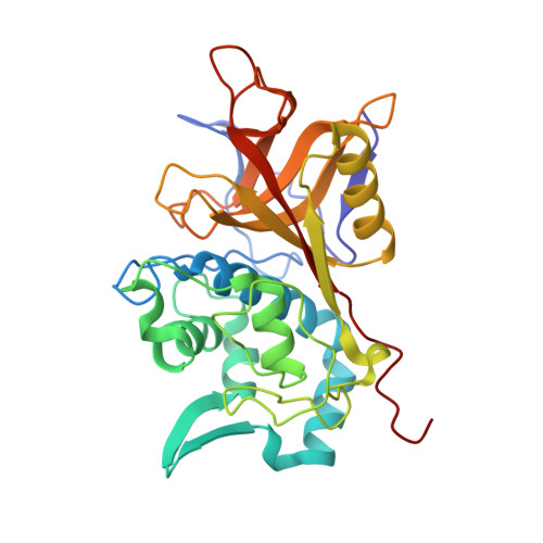
2AU1

Crystal Structure of group A Streptococcus MAC-1 orthorhombic form


Experimental Data Snapshot

  • Method: X-RAY DIFFRACTION
  • Resolution: 2.40 Å
  • R-Value Free: 0.266 
  • R-Value Work: 0.216 
  • R-Value Observed: 0.217 

wwPDB Validation   3D Report Full Report


This is version 1.3 of the entry. See complete history


Literature

Crystal structure of group a streptococcus mac-1: insight into dimer-mediated specificity for recognition of human IgG.

Agniswamy, J.Nagiec, M.J.Liu, M.Schuck, P.Musser, J.M.Sun, P.D.

(2006) Structure 14: 225-235

  • DOI: https://doi.org/10.1016/j.str.2005.10.012
  • Primary Citation of Related Structures:  
    2AU1, 2AVW

  • PubMed Abstract: 

    Group A Streptococcus secretes cysteine proteases named Mac-1 and Mac-2 that mediate host immune evasion by targeting both IgG and Fc receptors. Here, we report the crystal structures of Mac-1 and its catalytically inactive C94A mutant in two different crystal forms. Despite the lack of sequence homology, Mac-1 adopts the canonical papain fold. Alanine mutations at the active site confirmed the critical residues involved in a papain-like catalytic mechanism. Mac-1 forms a symmetric dimer in both crystal forms and displays the unique dimer interface among papain superfamily members. Mutations at the dimer interface resulted in a significant reduction in IgG binding and catalysis, suggesting that the dimer contributes to both IgG specificity and enzyme cooperativity. A tunnel observed at the dimer interface constitutes a target for designing potential Mac-1-specific antimicrobial agents. The structures also offer insight into the functional difference between Mac-1 and Mac-2.


  • Organizational Affiliation

    Structural Immunology Section, Laboratory of Immunogenetics, National Institute of Allergy and Infectious Diseases, National Institutes of Health, 12441 Parklawn Drive, Rockville, Maryland 20852, USA.


Macromolecules
Find similar proteins by:  (by identity cutoff)  |  3D Structure
Entity ID: 1
MoleculeChains Sequence LengthOrganismDetailsImage
IgG-degrading protease292Streptococcus pyogenes MGAS5005Mutation(s): 0 
UniProt
Find proteins for Q7DAM2 (Streptococcus pyogenes serotype M1)
Explore Q7DAM2 
Go to UniProtKB:  Q7DAM2
Entity Groups  
Sequence Clusters30% Identity50% Identity70% Identity90% Identity95% Identity100% Identity
UniProt GroupQ7DAM2
Sequence Annotations
Expand
  • Reference Sequence
Small Molecules
Ligands 1 Unique
IDChains Name / Formula / InChI Key2D Diagram3D Interactions
BME
Query on BME

Download Ideal Coordinates CCD File 
B [auth A]BETA-MERCAPTOETHANOL
C2 H6 O S
DGVVWUTYPXICAM-UHFFFAOYSA-N
Experimental Data & Validation

Experimental Data

  • Method: X-RAY DIFFRACTION
  • Resolution: 2.40 Å
  • R-Value Free: 0.266 
  • R-Value Work: 0.216 
  • R-Value Observed: 0.217 
  • Space Group: P 21 21 2
Unit Cell:
Length ( Å )Angle ( ˚ )
a = 63.865α = 90
b = 87.175β = 90
c = 57.603γ = 90
Software Package:
Software NamePurpose
HKL-2000data collection
SCALEPACKdata scaling
CNSrefinement
HKL-2000data reduction
CNSphasing

Structure Validation

View Full Validation Report


Entry History 

Deposition Data

Revision History  (Full details and data files)

  • Version 1.0: 2006-02-28
    Type: Initial release
  • Version 1.1: 2008-05-01
    Changes: Version format compliance
  • Version 1.2: 2011-07-13
    Changes: Source and taxonomy, Version format compliance
  • Version 1.3: 2024-02-14
    Changes: Data collection, Database references, Derived calculations