Refined structure of alpha-lytic protease at 1.7 A resolution. Analysis of hydrogen bonding and solvent structure.
Fujinaga, M., Delbaere, L.T., Brayer, G.D., James, M.N.(1985) J Mol Biol 184: 479-502
- PubMed: 3900416
- DOI: https://doi.org/10.1016/0022-2836(85)90296-7
- Primary Citation of Related Structures:
2ALP - PubMed Abstract:
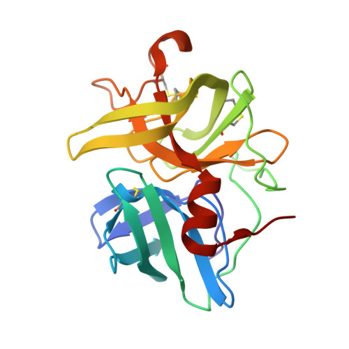
The structure of alpha-lytic protease, a serine protease produced by the bacterium Lysobacter enzymogenes, has been refined at 1.7 A resolution. The conventional R-factor is 0.131 for the 14,996 reflections between 8 and 1.7 A resolution with I greater than or equal to 2 sigma (I). The model consists of 1391 protein atoms, two sulfate ions and 156 water molecules. The overall root-meansquare error is estimated to be about 0.14 A. The refined structure was compared with homologous enzymes alpha-chymotrypsin and Streptomyces griseus protease A and B. A new sequence numbering was derived based on the alignment of these structures. The comparison showed that the greatest structural homology is around the active site residues Asp102, His57 and Ser195, and that basic folding pathways are maintained despite chemical changes in the hydrophobic cores. The hydrogen bonds in the structure were tabulated and the distances and angles of interaction are similar to those found in small molecules. The analysis also revealed the presence of close intraresidue interactions. There are only a few direct intermolecular hydrogen bonds. Most intermolecular interactions involve bridging solvent molecules. The structural importance of hydrogen bonds involving the side-chain of Asx residues is discussed. All the negatively charged groups have a counterion nearby, while the excess positively charged groups are exposed to the solvent. One of the sulfate ions is located near the active site, whereas the other is close to the N terminus. Of the 156 water molecules, only seven are not involved in a hydrogen bond. Six of these have polar groups nearby, while the remaining one is in very weak density. There are nine internal water molecules, consisting of two monomers, two dimers and one trimer. No significant second shell of solvent is observed.