2ADW

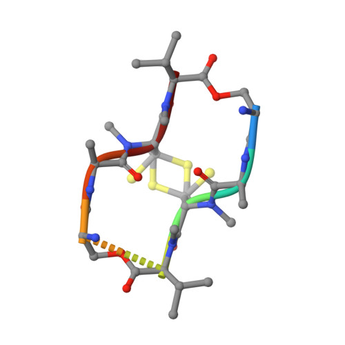
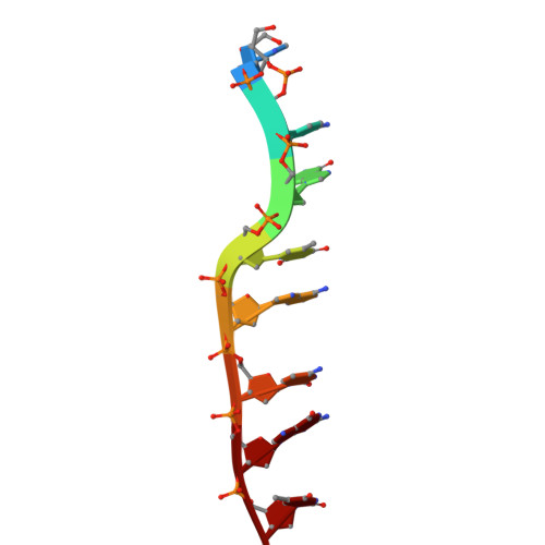
Crystal structure of Echinomycin-(ACGTACGT)2 solved by SAD


Experimental Data Snapshot

  • Method: X-RAY DIFFRACTION
  • Resolution: 1.60 Å
  • R-Value Free: 0.221 
  • R-Value Observed: 0.180 

wwPDB Validation   3D Report Full Report


This is version 1.5 of the entry. See complete history


Literature

Serendipitous Sad Phasing of an Echinomycin-(Acgtacgt)2 Bisintercalation Complex.

Cuesta-Seijo, J.A.Weiss, M.S.Sheldrick, G.M.

(2006) Acta Crystallogr D Biol Crystallogr 62: 417

  • DOI: https://doi.org/10.1107/S0907444906003763
  • Primary Citation of Related Structures:  
    2ADW

  • PubMed Abstract: 

    A new crystal form was obtained for the complex between (ACGTACGT)2 and echinomycin and X-ray data were collected to 1.6 A. The structure was phased by the SAD method based on a single unexpected anomalous scatterer that could be identified as a mixture of nickel and zinc by measurements of the anomalous scattering at different wavelengths. This cation is coordinated by two guanines from two different duplexes and four water molecules. The structure resembles previously reported crystal structures of DNA-echinomycin complexes, except that three of the eight base pairs flanking the echinomycin bisintercalator sites have the Watson-Crick rather than the Hoogsteen configuration. Hoogsteen binding was found for the corresponding base pairs of the crystallographically independent duplex, indicating that the two configurations are very close in energy.


  • Organizational Affiliation

    Department of Structural Chemistry, University of Göttingen, Tammannstrasse 4, D37077 Göttingen, Germany.


Macromolecules

Find similar proteins by:  Sequence   |   3D Structure  

Entity ID: 2
MoleculeChains Sequence LengthOrganismDetailsImage
ECHINOMYCINE [auth H],
F [auth I],
G [auth J],
H [auth K]
8Streptomyces echinatusMutation(s): 2 
Sequence Annotations
Expand
  • Reference Sequence

Find similar nucleic acids by:  Sequence   |   3D Structure  

Entity ID: 1
MoleculeChains LengthOrganismImage
5'-D(*AP*CP*GP*TP*AP*CP*GP*T)-3'A,
B [auth C],
C [auth E],
D [auth G]
8N/A
Sequence Annotations
Expand
  • Reference Sequence
Small Molecules
Ligands 4 Unique
IDChains Name / Formula / InChI Key2D Diagram3D Interactions
MES
Query on MES

Download Ideal Coordinates CCD File 
I [auth C]2-(N-MORPHOLINO)-ETHANESULFONIC ACID
C6 H13 N O4 S
SXGZJKUKBWWHRA-UHFFFAOYSA-N
QUI
Query on QUI

Download Ideal Coordinates CCD File 
L [auth H]
M [auth H]
N [auth I]
O [auth I]
P [auth J]
L [auth H],
M [auth H],
N [auth I],
O [auth I],
P [auth J],
Q [auth J],
R [auth K],
S [auth K]
2-CARBOXYQUINOXALINE
C9 H6 N2 O2
UPUZGXILYFKSGE-UHFFFAOYSA-N
ZN
Query on ZN

Download Ideal Coordinates CCD File 
K [auth E]ZINC ION
Zn
PTFCDOFLOPIGGS-UHFFFAOYSA-N
NI
Query on NI

Download Ideal Coordinates CCD File 
J [auth E]NICKEL (II) ION
Ni
VEQPNABPJHWNSG-UHFFFAOYSA-N
Modified Residues  1 Unique
IDChains TypeFormula2D DiagramParent
MVA
Query on MVA
E [auth H],
F [auth I],
G [auth J],
H [auth K]
L-PEPTIDE LINKINGC6 H13 N O2VAL
Experimental Data & Validation

Experimental Data

  • Method: X-RAY DIFFRACTION
  • Resolution: 1.60 Å
  • R-Value Free: 0.221 
  • R-Value Observed: 0.180 
  • Space Group: P 42 21 2
Unit Cell:
Length ( Å )Angle ( ˚ )
a = 80.904α = 90
b = 80.904β = 90
c = 48.194γ = 90
Software Package:
Software NamePurpose
SHELXDphasing
SHELXEmodel building
SHELXL-97refinement
HKL-2000data reduction
XDSdata scaling

Structure Validation

View Full Validation Report


Entry History 

Deposition Data

Revision History  (Full details and data files)

  • Version 1.0: 2006-03-28
    Type: Initial release
  • Version 1.1: 2011-06-14
    Changes: Version format compliance
  • Version 1.2: 2011-07-13
    Changes: Version format compliance
  • Version 1.3: 2011-07-27
    Changes: Atomic model, Database references, Derived calculations, Non-polymer description, Structure summary
  • Version 1.4: 2012-12-12
    Changes: Other
  • Version 1.5: 2020-09-09
    Changes: Advisory, Database references, Derived calculations, Structure summary