2A8K

Structural and Mutational Studies of the Catalytic Domain of Colicin E5a tRNA-Specific Ribonuclease


Experimental Data Snapshot

  • Method: X-RAY DIFFRACTION
  • Resolution: 1.50 Å
  • R-Value Free: 0.234 
  • R-Value Work: 0.217 

wwPDB Validation   3D Report Full Report


This is version 1.3 of the entry. See complete history


Literature

Structural and mutational studies of the catalytic domain of colicin E5: A tRNA-specific ribonuclease

Lin, Y.L.Elias, Y.Huang, R.H.

(2005) Biochemistry 44: 10494-10500

  • DOI: https://doi.org/10.1021/bi050749s
  • Primary Citation of Related Structures:  
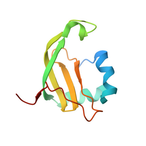
    2A8K

  • PubMed Abstract: 

    Colicin E5 specifically cleaves four tRNAs in Escherichia coli that contain the modified nucleotide queuosine (Q) at the wobble position, thereby preventing protein synthesis and ultimately resulting in cell death. Here, the crystal structure of the catalytic domain of colicin E5 (E5-CRD) from E. coli was determined at 1.5 A resolution. Unexpectedly, E5-CRD adopts a core folding with a four-stranded beta-sheet packed against an alpha-helix, seen in the well-studied ribonuclease T1 despite a lack of sequence similarity. Beyond the core catalytic domain, an N-terminal helix, a C-terminal beta-strand and loop, and an extended internal loop constitute an RNA binding cleft. Mutational analysis identified five amino acids that were important for tRNA substrate binding and cleavage by E5-CRD. The structure, together with the mutational study, allows us to propose a model of colicin E5-tRNA interactions, suggesting the molecular basis of tRNA substrate recognition and the mechanism of tRNA cleavage by colicin E5.


  • Organizational Affiliation

    Department of Chemistry, University of Illinois, 600 South Mathews Avenue, Urbana, Illinois 61801, USA.


Macromolecules
Find similar proteins by:  (by identity cutoff)  |  3D Structure
Entity ID: 1
MoleculeChains Sequence LengthOrganismDetailsImage
Colicin E5
A, B, C, D
108Escherichia coliMutation(s): 0 
Gene Names: col
EC: 3.1
UniProt
Find proteins for P18000 (Escherichia coli)
Explore P18000 
Go to UniProtKB:  P18000
Entity Groups  
Sequence Clusters30% Identity50% Identity70% Identity90% Identity95% Identity100% Identity
UniProt GroupP18000
Sequence Annotations
Expand
  • Reference Sequence
Experimental Data & Validation

Experimental Data

  • Method: X-RAY DIFFRACTION
  • Resolution: 1.50 Å
  • R-Value Free: 0.234 
  • R-Value Work: 0.217 
  • Space Group: P 31
Unit Cell:
Length ( Å )Angle ( ˚ )
a = 65.22α = 90
b = 65.22β = 90
c = 100.74γ = 120
Software Package:
Software NamePurpose
HKL-2000data collection
SCALEPACKdata scaling
SOLVEphasing
CNSrefinement
HKL-2000data reduction

Structure Validation

View Full Validation Report


Entry History 

Deposition Data

Revision History  (Full details and data files)

  • Version 1.0: 2005-09-20
    Type: Initial release
  • Version 1.1: 2008-04-30
    Changes: Version format compliance
  • Version 1.2: 2011-07-13
    Changes: Version format compliance
  • Version 1.3: 2024-02-14
    Changes: Data collection, Database references, Derived calculations