2A7X

Crystal Structure of A Pantothenate synthetase complexed with AMP


Experimental Data Snapshot

  • Method: X-RAY DIFFRACTION
  • Resolution: 1.70 Å
  • R-Value Free: 0.175 
  • R-Value Work: 0.148 
  • R-Value Observed: 0.150 

wwPDB Validation   3D Report Full Report


Ligand Structure Quality Assessment 


This is version 1.4 of the entry. See complete history


Literature

Crystal Structure of the Pantothenate Synthetase from Mycobacterium tuberculosis, Snapshots of the Enzyme in Action.

Wang, S.Eisenberg, D.

(2006) Biochemistry 45: 1554-1561

  • DOI: https://doi.org/10.1021/bi051873e
  • Primary Citation of Related Structures:  
    2A7X, 2A84, 2A86, 2A88

  • PubMed Abstract: 

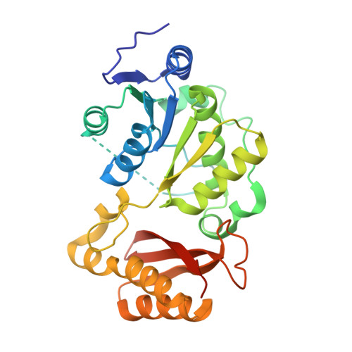
    Pantothenate synthetase (PS) from Mycobacterium tuberculosis represents a potential target for antituberculosis drugs. PS catalyzes the ATP-dependent condensation of pantoate and beta-alanine to form pantothenate. Previously, we determined the crystal structure of PS from M. tuberculosis and its complexes with AMPCPP, pantoate, and pantoyl adenylate. Here, we describe the crystal structure of this enzyme complexed with AMP and its last substrate, beta-alanine, and show that the phosphate group of AMP serves as an anchor for the binding of beta-alanine. This structure confirms that binding of beta-alanine in the active site cavity can occur only after formation of the pantoyl adenylate intermediate. A new crystal form was also obtained; it displays the flexible wall of the active site cavity in a conformation incapable of binding pantoate. Soaking of this crystal form with ATP and pantoate gives a fully occupied complex of PS with ATP. Crystal structures of these complexes with substrates, the reaction intermediate, and the reaction product AMP provide a step-by-step view of the PS-catalyzed reaction. A detailed reaction mechanism and its implications for inhibitor design are discussed.


  • Organizational Affiliation

    Public Health Research Institute, 225 Warren Street, Newark, New Jersey 07103, USA. shuishu@phri.org


Macromolecules
Find similar proteins by:  (by identity cutoff)  |  3D Structure
Entity ID: 1
MoleculeChains Sequence LengthOrganismDetailsImage
Pantoate-beta-alanine ligase300Mycobacterium tuberculosisMutation(s): 2 
Gene Names: panC
EC: 6.3.2.1
UniProt
Find proteins for P9WIL5 (Mycobacterium tuberculosis (strain ATCC 25618 / H37Rv))
Explore P9WIL5 
Go to UniProtKB:  P9WIL5
Entity Groups  
Sequence Clusters30% Identity50% Identity70% Identity90% Identity95% Identity100% Identity
UniProt GroupP9WIL5
Sequence Annotations
Expand
  • Reference Sequence
Experimental Data & Validation

Experimental Data

  • Method: X-RAY DIFFRACTION
  • Resolution: 1.70 Å
  • R-Value Free: 0.175 
  • R-Value Work: 0.148 
  • R-Value Observed: 0.150 
  • Space Group: C 1 2 1
Unit Cell:
Length ( Å )Angle ( ˚ )
a = 120.262α = 90
b = 44.944β = 125.56
c = 82.236γ = 90
Software Package:
Software NamePurpose
REFMACrefinement
DENZOdata reduction
SCALEPACKdata scaling
AMoREphasing

Structure Validation

View Full Validation Report


Ligand Structure Quality Assessment 


Entry History 

Deposition Data

Revision History  (Full details and data files)

  • Version 1.0: 2006-02-21
    Type: Initial release
  • Version 1.1: 2008-04-30
    Changes: Version format compliance
  • Version 1.2: 2011-07-13
    Changes: Advisory, Version format compliance
  • Version 1.3: 2021-10-20
    Changes: Database references, Derived calculations
  • Version 1.4: 2023-08-23
    Changes: Data collection, Refinement description