2ZXG

Aminopeptidase N complexed with the aminophosphinic inhibitor of PL250, a transition state analogue


Experimental Data Snapshot

  • Method: X-RAY DIFFRACTION
  • Resolution: 1.55 Å
  • R-Value Free: 0.195 
  • R-Value Work: 0.178 
  • R-Value Observed: 0.179 

wwPDB Validation   3D Report Full Report


Ligand Structure Quality Assessment 


This is version 1.2 of the entry. See complete history


Literature

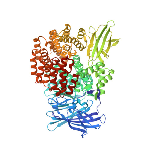
Structure of aminopeptidase N from Escherichia coli complexed with the transition-state analogue aminophosphinic inhibitor PL250

Fournie-Zaluski, M.C.Poras, H.Roques, B.P.Nakajima, Y.Ito, K.Yoshimoto, T.

(2009) Acta Crystallogr D Biol Crystallogr 65: 814-822

  • DOI: https://doi.org/10.1107/S090744490901779X
  • Primary Citation of Related Structures:  
    2ZXG

  • PubMed Abstract: 

    Aminopeptidase N (APN; EC 3.4.11.2) purified from Escherichia coli has been crystallized with the optically pure aminophosphinic inhibitor PL250, H(3)N(+)-CH(CH(3))-P(O)(OH)-CH(2)-CH(CH(2)Ph)-CONH-CH(CH(2)Ph)CO(2)(-), which mimics the transition state of the hydrolysis reaction. PL250 inhibits APN with a K(i) of 1.5-2.2 nM and its three-dimensional structure in complex with E. coli APN showed its interaction with the S(1), S'(1) and S'(2) subsites of the catalytic site. In this structure, the Zn ion was shown to be pentacoordinated by His297, His301 and Glu320 of APN and the two O atoms of the phosphinic moiety of PL250. One of these O atoms is also involved in a hydrogen bond to Tyr381, supporting the proposed role of this amino acid in the stabilization of the transition state of the enzymatic process. The strength of the phosphinic zinc binding and the occupancy of the S'(2) subsite account for the 100-fold increase in affinity of PL250 compared with the dipeptide-derived inhibitor bestatin (K(i) = 4.1 x 10(-6) M). Accordingly, the removal of the C-terminal phenylalanine of PL250 resulted in a large decrease in affinity (K(i) = 2.17 x 10(-7) M). Furthermore, it was observed that the C-terminal carboxyl group of the inhibitor makes no direct interactions with the amino acids of the APN active site. Interestingly, PL250 exhibits the same inhibitory potency for E. coli APN and for mammalian enzymes, suggesting that the structure of the complex could be used as a template for the rational design of various human APN inhibitors needed to study the role of this aminopeptidase in various pathologies.


  • Organizational Affiliation

    Pharmaleads, France.


Macromolecules
Find similar proteins by:  (by identity cutoff)  |  3D Structure
Entity ID: 1
MoleculeChains Sequence LengthOrganismDetailsImage
Aminopeptidase N870Escherichia coli K-12Mutation(s): 0 
Gene Names: pepN
EC: 3.4.11.2
UniProt
Find proteins for P04825 (Escherichia coli (strain K12))
Explore P04825 
Go to UniProtKB:  P04825
Entity Groups  
Sequence Clusters30% Identity50% Identity70% Identity90% Identity95% Identity100% Identity
UniProt GroupP04825
Sequence Annotations
Expand
  • Reference Sequence
Small Molecules
Ligands 4 Unique
IDChains Name / Formula / InChI Key2D Diagram3D Interactions
S23
Query on S23

Download Ideal Coordinates CCD File 
C [auth A]N-{(2S)-3-[(1R)-1-aminoethyl](hydroxy)phosphoryl-2-benzylpropanoyl}-L-phenylalanine
C21 H27 N2 O5 P
MMWNOZXGYRPSCJ-LZQZEXGQSA-N
SO4
Query on SO4

Download Ideal Coordinates CCD File 
D [auth A]SULFATE ION
O4 S
QAOWNCQODCNURD-UHFFFAOYSA-L
GOL
Query on GOL

Download Ideal Coordinates CCD File 
E [auth A]
F [auth A]
G [auth A]
H [auth A]
I [auth A]
E [auth A],
F [auth A],
G [auth A],
H [auth A],
I [auth A],
J [auth A],
K [auth A]
GLYCEROL
C3 H8 O3
PEDCQBHIVMGVHV-UHFFFAOYSA-N
ZN
Query on ZN

Download Ideal Coordinates CCD File 
B [auth A]ZINC ION
Zn
PTFCDOFLOPIGGS-UHFFFAOYSA-N
Binding Affinity Annotations 
IDSourceBinding Affinity
S23 PDBBind:  2ZXG Ki: 1.5 (nM) from 1 assay(s)
Binding MOAD:  2ZXG Ki: 790 (nM) from 1 assay(s)
Experimental Data & Validation

Experimental Data

  • Method: X-RAY DIFFRACTION
  • Resolution: 1.55 Å
  • R-Value Free: 0.195 
  • R-Value Work: 0.178 
  • R-Value Observed: 0.179 
  • Space Group: P 31 2 1
Unit Cell:
Length ( Å )Angle ( ˚ )
a = 120.7α = 90
b = 120.7β = 90
c = 171γ = 120
Software Package:
Software NamePurpose
REFMACrefinement
ADSCdata collection
HKL-2000data reduction
HKL-2000data scaling
CNSphasing

Structure Validation

View Full Validation Report


Ligand Structure Quality Assessment 


Entry History 

Deposition Data

Revision History  (Full details and data files)

  • Version 1.0: 2009-08-25
    Type: Initial release
  • Version 1.1: 2011-07-13
    Changes: Non-polymer description, Version format compliance
  • Version 1.2: 2023-11-01
    Changes: Data collection, Database references, Derived calculations, Refinement description