2ZWI

Crystal structure of alpha/beta-Galactoside alpha-2,3-Sialyltransferase from a Luminous Marine Bacterium, <I>Photobacterium phosphoreum</I>


Experimental Data Snapshot

  • Method: X-RAY DIFFRACTION
  • Resolution: 2.01 Å
  • R-Value Free: 0.228 
  • R-Value Work: 0.173 
  • R-Value Observed: 0.176 

wwPDB Validation   3D Report Full Report


Ligand Structure Quality Assessment 


This is version 1.2 of the entry. See complete history


Literature

Crystal structure of alpha/beta-galactoside alpha2,3-sialyltransferase from a luminous marine bacterium, Photobacterium phosphoreum

Iwatani, T.Okino, N.Sakakura, M.Kajiwara, H.Takakura, Y.Kimura, M.Ito, M.Yamamoto, T.Kakuta, Y.

(2009) FEBS Lett 583: 2083-2087

  • DOI: https://doi.org/10.1016/j.febslet.2009.05.032
  • Primary Citation of Related Structures:  
    2ZWI

  • PubMed Abstract: 

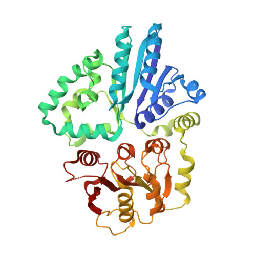
    Alpha/beta-galactoside alpha2,3-sialyltransferase produced by Photobacterium phosphoreum JT-ISH-467 is a unique enzyme that catalyzes the transfer of N-acetylneuraminic acid residue from cytidine monophosphate N-acetylneuraminic acid to acceptor carbohydrate groups. The enzyme recognizes both mono- and di-saccharides as acceptor substrates, and can transfer Neu5Ac to both alpha-galactoside and beta-galactoside, efficiently. To elucidate the structural basis for the broad acceptor substrate specificity, we determined the crystal structure of the alpha2,3-sialyltransferase in complex with CMP. The overall structure belongs to the glycosyltransferase-B structural group. We could model a reasonable active conformation structure based on the crystal structure. The predicted structure suggested that the broad substrate specificity could be attributed to the wider entrance of the acceptor substrate binding site.


  • Organizational Affiliation

    Laboratory of Structural Biology, Graduate School of Systems Life Sciences, Kyushu University, Fukuoka 812-8581, Japan.


Macromolecules
Find similar proteins by:  (by identity cutoff)  |  3D Structure
Entity ID: 1
MoleculeChains Sequence LengthOrganismDetailsImage
Alpha-/beta-galactoside alpha-2,3-sialyltransferase
A, B
373Photobacterium phosphoreumMutation(s): 0 
Gene Names: pst3-467
EC: 2.4.99.4
UniProt
Find proteins for A5LHX0 (Photobacterium phosphoreum)
Explore A5LHX0 
Go to UniProtKB:  A5LHX0
Entity Groups  
Sequence Clusters30% Identity50% Identity70% Identity90% Identity95% Identity100% Identity
UniProt GroupA5LHX0
Sequence Annotations
Expand
  • Reference Sequence
Small Molecules
Ligands 4 Unique
IDChains Name / Formula / InChI Key2D Diagram3D Interactions
C5P
Query on C5P

Download Ideal Coordinates CCD File 
C [auth A],
K [auth B]
CYTIDINE-5'-MONOPHOSPHATE
C9 H14 N3 O8 P
IERHLVCPSMICTF-XVFCMESISA-N
GOL
Query on GOL

Download Ideal Coordinates CCD File 
E [auth A]
F [auth A]
G [auth A]
H [auth A]
M [auth B]
E [auth A],
F [auth A],
G [auth A],
H [auth A],
M [auth B],
N [auth B],
O [auth B],
P [auth B],
Q [auth B]
GLYCEROL
C3 H8 O3
PEDCQBHIVMGVHV-UHFFFAOYSA-N
ZN
Query on ZN

Download Ideal Coordinates CCD File 
D [auth A],
L [auth B]
ZINC ION
Zn
PTFCDOFLOPIGGS-UHFFFAOYSA-N
CL
Query on CL

Download Ideal Coordinates CCD File 
I [auth A],
J [auth A],
R [auth B],
S [auth B]
CHLORIDE ION
Cl
VEXZGXHMUGYJMC-UHFFFAOYSA-M
Experimental Data & Validation

Experimental Data

  • Method: X-RAY DIFFRACTION
  • Resolution: 2.01 Å
  • R-Value Free: 0.228 
  • R-Value Work: 0.173 
  • R-Value Observed: 0.176 
  • Space Group: P 1
Unit Cell:
Length ( Å )Angle ( ˚ )
a = 54.429α = 90.06
b = 57.827β = 106.76
c = 59.246γ = 103.2
Software Package:
Software NamePurpose
REFMACrefinement
HKL-2000data collection
HKL-2000data scaling
MOLREPphasing

Structure Validation

View Full Validation Report


Ligand Structure Quality Assessment 


Entry History 

Deposition Data

Revision History  (Full details and data files)

  • Version 1.0: 2009-06-09
    Type: Initial release
  • Version 1.1: 2011-07-13
    Changes: Non-polymer description, Version format compliance
  • Version 1.2: 2023-11-01
    Changes: Data collection, Database references, Derived calculations, Refinement description