


Crystal structure of pyridoxine biosynthesis protein from Thermus thermophilus HB8
Manzoku, M., Ebihara, A., Fujimoto, Y., Yokoyama, S., Kuramitsu, S.To be published.
Experimental Data Snapshot
wwPDB Validation 3D Report Full Report

Entity ID: 1 | |||||
|---|---|---|---|---|---|
| Molecule | Chains | Sequence Length | Organism | Details | Image |
| Pyridoxal biosynthesis lyase pdxS | 297 | Thermus thermophilus HB8 | Mutation(s): 0 Gene Names: pdxS EC: 4 | 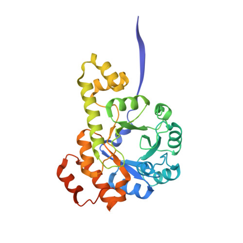 | |
UniProt | |||||
Find proteins for Q5SKD9 (Thermus thermophilus (strain ATCC 27634 / DSM 579 / HB8)) Explore Q5SKD9 Go to UniProtKB: Q5SKD9 | |||||
Entity Groups | |||||
| Sequence Clusters | 30% Identity50% Identity70% Identity90% Identity95% Identity100% Identity | ||||
| UniProt Group | Q5SKD9 | ||||
Sequence AnnotationsExpand | |||||
| |||||
| Ligands 1 Unique | |||||
|---|---|---|---|---|---|
| ID | Chains | Name / Formula / InChI Key | 2D Diagram | 3D Interactions | |
| MPD Query on MPD | E [auth A], F [auth B], G [auth C], H [auth D] | (4S)-2-METHYL-2,4-PENTANEDIOL C6 H14 O2 SVTBMSDMJJWYQN-YFKPBYRVSA-N |  | ||
| Length ( Å ) | Angle ( ˚ ) |
|---|---|
| a = 180.97 | α = 90 |
| b = 180.97 | β = 90 |
| c = 100.75 | γ = 120 |
| Software Name | Purpose |
|---|---|
| CNS | refinement |
| HKL-2000 | data collection |
| HKL-2000 | data reduction |
| HKL-2000 | data scaling |
| SOLVE | phasing |
RCSB PDB (citation) is hosted by
RCSB PDB is a member of the

