2ZBO

Crystal structure of low-redox-potential cytochrom c6 from brown alga Hizikia fusiformis at 1.6 A resolution


Experimental Data Snapshot

  • Method: X-RAY DIFFRACTION
  • Resolution: 1.60 Å
  • R-Value Free: 0.209 
  • R-Value Work: 0.184 
  • R-Value Observed: 0.185 

wwPDB Validation   3D Report Full Report


Ligand Structure Quality Assessment 


This is version 1.2 of the entry. See complete history


Literature

Cloning, expression and purification of cytochrome c(6) from the brown alga Hizikia fusiformis and complete X-ray diffraction analysis of the structure

Akazaki, H.Kawai, F.Chida, H.Matsumoto, Y.Hirayama, M.Hoshikawa, K.Unzai, S.Hakamata, W.Nishio, T.Park, S.-Y.Oku, T.

(2008) Acta Crystallogr Sect F Struct Biol Cryst Commun 64: 674-680

  • DOI: https://doi.org/10.1107/S1744309108017752
  • Primary Citation of Related Structures:  
    2ZBO

  • PubMed Abstract: 

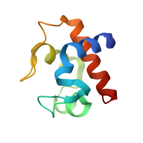
    The primary sequence of cytochrome c(6) from the brown alga Hizikia fusiformis has been determined by cDNA cloning and the crystal structure has been solved at 1.6 A resolution. The crystal belonged to the tetragonal space group P4(1)2(1)2, with unit-cell parameters a = b = 84.58, c = 232.91 A and six molecules per asymmetric unit. The genome code, amino-acid sequence and crystal structure of H. fusiformis cytochrome c(6) were most similar to those of red algal cytochrome c(6). These results support the hypothesis that brown algae acquired their chloroplasts via secondary endosymbiosis involving a red algal endosymbiont and a eukaryote host.


  • Organizational Affiliation

    Bio-organic Chemistry Laboratory, Graduate School of Bioresource Sciences, Nihon University, Kameino 1866, Fujisawa-shi, Kanagawa 252-8510, Japan.


Macromolecules
Find similar proteins by:  (by identity cutoff)  |  3D Structure
Entity ID: 1
MoleculeChains Sequence LengthOrganismDetailsImage
Cytochrome c686Sargassum fusiformeMutation(s): 0 
Gene Names: petJ
UniProt
Find proteins for Q76FN8 (Sargassum fusiforme)
Explore Q76FN8 
Go to UniProtKB:  Q76FN8
Entity Groups  
Sequence Clusters30% Identity50% Identity70% Identity90% Identity95% Identity100% Identity
UniProt GroupQ76FN8
Sequence Annotations
Expand
  • Reference Sequence
Small Molecules
Ligands 2 Unique
IDChains Name / Formula / InChI Key2D Diagram3D Interactions
HEM
Query on HEM

Download Ideal Coordinates CCD File 
G [auth A]
I [auth C]
J [auth E]
K [auth G]
M [auth I]
G [auth A],
I [auth C],
J [auth E],
K [auth G],
M [auth I],
O [auth K]
PROTOPORPHYRIN IX CONTAINING FE
C34 H32 Fe N4 O4
KABFMIBPWCXCRK-RGGAHWMASA-L
SO4
Query on SO4

Download Ideal Coordinates CCD File 
H [auth A],
L [auth G],
N [auth I],
P [auth K]
SULFATE ION
O4 S
QAOWNCQODCNURD-UHFFFAOYSA-L
Experimental Data & Validation

Experimental Data

  • Method: X-RAY DIFFRACTION
  • Resolution: 1.60 Å
  • R-Value Free: 0.209 
  • R-Value Work: 0.184 
  • R-Value Observed: 0.185 
  • Space Group: P 41 21 2
Unit Cell:
Length ( Å )Angle ( ˚ )
a = 84.578α = 90
b = 84.578β = 90
c = 232.911γ = 90
Software Package:
Software NamePurpose
REFMACrefinement
HKL-2000data reduction
SCALEPACKdata scaling
MOLREPphasing

Structure Validation

View Full Validation Report


Ligand Structure Quality Assessment 


Entry History 

Revision History  (Full details and data files)

  • Version 1.0: 2008-09-30
    Type: Initial release
  • Version 1.1: 2011-07-13
    Changes: Version format compliance
  • Version 1.2: 2023-11-01
    Changes: Data collection, Database references, Derived calculations, Refinement description