Crystal Structures of Pseudomonas Aeruginosa Gim-1: Active-Site Plasticity in Metallo-Beta-Lactamases.
Borra, P.S., Samuelsen, O., Spencer, J., Walsh, T.R., Lorentzen, M.S., Leiros, H.-K.S.(2013) Antimicrob Agents Chemother 57: 848
- PubMed: 23208706
- DOI: https://doi.org/10.1128/AAC.02227-12
- Primary Citation of Related Structures:
2YNT, 2YNU, 2YNV, 2YNW - PubMed Abstract:
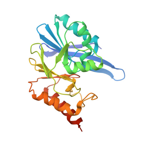
Metallo-β-lactamases (MBLs) have rapidly disseminated worldwide among clinically important Gram-negative bacteria and have challenged the therapeutic use of β-lactam antibiotics, particularly carbapenems. The bla(GIM-1) gene, encoding one such enzyme, was first discovered in a Pseudomonas aeruginosa isolate from 2002 and has more recently been reported in Enterobacteriaceae. Here, we present crystal structures of GIM-1 in the apo-zinc (metal-free), mono-zinc (where Cys221 was found to be oxidized), and di-zinc forms, providing nine independently refined views of the enzyme. GIM-1 is distinguished from related MBLs in possessing a narrower active-site groove defined by aromatic side chains (Trp228 and Tyr233) at positions normally occupied by hydrophilic residues in other MBLs. Our structures reveal considerable flexibility in two loops (loop 1, residues 60 to 66; loop 2, residues 223 to 242) adjacent to the active site, with open and closed conformations defined by alternative hydrogen-bonding patterns involving Trp228. We suggest that this capacity for rearrangement permits GIM-1 to hydrolyze a wide range of β-lactams in spite of possessing a more constrained active site. Our results highlight the structural diversity within the MBL enzyme family.
Organizational Affiliation:
Research Group for Host-Microbe Interactions, Department of Medical Biology, Faculty of Health Sciences, University of Tromsø, Tromsø, Norway.