2Y75

The Structure of CymR (YrzC) the Global Cysteine Regulator of B. subtilis


Experimental Data Snapshot

  • Method: X-RAY DIFFRACTION
  • Resolution: 2.00 Å
  • R-Value Free: 0.214 
  • R-Value Work: 0.187 
  • R-Value Observed: 0.188 

wwPDB Validation   3D Report Full Report


This is version 1.1 of the entry. See complete history


Literature

Insights Into the Rrf2 Repressor Family - the Structure of Cymr, the Global Cysteine Regulator of Bacillus Subtilis.

Shepard, W.Soutourina, O.Courtois, E.England, P.Haouz, A.Martin-Verstraete, I.

(2011) FEBS J 278: 2689

  • DOI: https://doi.org/10.1111/j.1742-4658.2011.08195.x
  • Primary Citation of Related Structures:  
    2Y75

  • PubMed Abstract: 

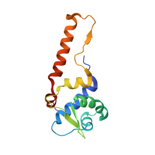
    The global regulator CymR represses the transcription of a large set of genes involved in cystine uptake and cysteine biosynthesis in Bacillus subtilis and Staphylococcus aureus. This repressor belongs to the widespread and poorly characterized Rrf2 family of regulators. The crystal structure of CymR from B. subtilis reveals a biologically active dimer, where each monomer folds into two tightly packed domains: a DNA-binding domain, which houses a winged helix-turn-helix (wHTH) motif; and a long dimerization domain, which places the wHTH motifs at the extremes. This architecture explains how these small regulators can span 23-27-bp DNA targets. The wHTH motif of CymR resembles those of the GntR superfamily of regulators, such as FadR and HutC. Superimposing the FadR wHTH motifs bound to their DNA fragments onto the wHTH motifs of the CymR dimer structure suggests that the DNA target and/or the protein must undergo some conformational changes upon binding. The CymR structure also hints at a possible location of the Fe-S centre associated with several Rrf2-type regulators.


  • Organizational Affiliation

    Synchrotron SOLEIL, L'Orme des Merisiers, Saint Aubin, BP48, Gif-sur-Yvette, France.


Macromolecules
Find similar proteins by:  (by identity cutoff)  |  3D Structure
Entity ID: 1
MoleculeChains Sequence LengthOrganismDetailsImage
HTH-TYPE TRANSCRIPTIONAL REGULATOR CYMR
A, B, C, D, E
A, B, C, D, E, F
129Bacillus subtilisMutation(s): 0 
UniProt
Find proteins for O34527 (Bacillus subtilis (strain 168))
Explore O34527 
Go to UniProtKB:  O34527
Entity Groups  
Sequence Clusters30% Identity50% Identity70% Identity90% Identity95% Identity100% Identity
UniProt GroupO34527
Sequence Annotations
Expand
  • Reference Sequence
Small Molecules
Ligands 2 Unique
IDChains Name / Formula / InChI Key2D Diagram3D Interactions
SO4
Query on SO4

Download Ideal Coordinates CCD File 
G [auth A]
H [auth A]
I [auth A]
J [auth B]
K [auth B]
G [auth A],
H [auth A],
I [auth A],
J [auth B],
K [auth B],
L [auth B],
N [auth C],
O [auth C],
P [auth D],
Q [auth D],
S [auth E],
T [auth E],
U [auth F],
V [auth F],
W [auth F],
Y [auth F]
SULFATE ION
O4 S
QAOWNCQODCNURD-UHFFFAOYSA-L
CL
Query on CL

Download Ideal Coordinates CCD File 
M [auth B],
R [auth D],
X [auth F]
CHLORIDE ION
Cl
VEXZGXHMUGYJMC-UHFFFAOYSA-M
Experimental Data & Validation

Experimental Data

  • Method: X-RAY DIFFRACTION
  • Resolution: 2.00 Å
  • R-Value Free: 0.214 
  • R-Value Work: 0.187 
  • R-Value Observed: 0.188 
  • Space Group: P 21 21 21
Unit Cell:
Length ( Å )Angle ( ˚ )
a = 98.7α = 90
b = 103.7β = 90
c = 106.53γ = 90
Software Package:
Software NamePurpose
BUSTERrefinement
XDSdata reduction
XDSdata scaling

Structure Validation

View Full Validation Report


Entry History 

Deposition Data

Revision History  (Full details and data files)

  • Version 1.0: 2011-08-03
    Type: Initial release
  • Version 1.1: 2019-05-08
    Changes: Data collection, Experimental preparation, Other