2XVW

Human serum albumin complexed with dansyl-L-arginine and myristic acid


Experimental Data Snapshot

  • Method: X-RAY DIFFRACTION
  • Resolution: 2.65 Å
  • R-Value Free: 0.252 
  • R-Value Work: 0.191 
  • R-Value Observed: 0.191 

wwPDB Validation   3D Report Full Report


Ligand Structure Quality Assessment 


This is version 1.4 of the entry. See complete history


Literature

Structural Basis of Binding of Fluorescent, Site-Specific Dansylated Amino Acids to Human Serum Albumin.

Ryan, A.J.Ghuman, J.Zunszain, P.A.Chung, C.W.Curry, S.

(2011) J Struct Biol 174: 84

  • DOI: https://doi.org/10.1016/j.jsb.2010.10.004
  • Primary Citation of Related Structures:  
    2XSI, 2XVQ, 2XVU, 2XVV, 2XVW, 2XW0, 2XW1

  • PubMed Abstract: 

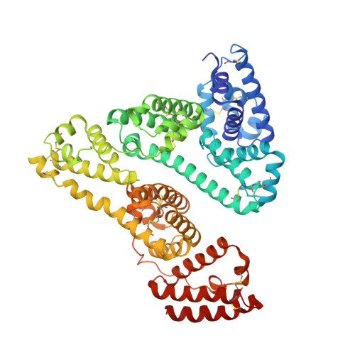
    Human serum albumin (HSA) has two primary binding sites for drug molecules. These sites selectively bind different dansylated amino acid compounds, which-due to their intrinsic fluorescence-have long been used as specific markers for the drug pockets on HSA. We present here the co-crystal structures of HSA in complex with six dansylated amino acids that are specific for either drug site 1 (dansyl-l-asparagine, dansyl-l-arginine, dansyl-l-glutamate) or drug site 2 (dansyl-l-norvaline, dansyl-l-phenylalanine, dansyl-l-sarcosine). Our results explain the structural basis of the site-specificity of different dansylated amino acids. They also show that fatty acid binding has only a modest effect on binding of dansylated amino acids to drug site 1 and identify the location of secondary binding sites.


  • Organizational Affiliation

    Biophysics Section, Blackett Laboratory, Imperial College, Exhibition Road, London SW7 2AZ, United Kingdom.


Macromolecules
Find similar proteins by:  (by identity cutoff)  |  3D Structure
Entity ID: 1
MoleculeChains Sequence LengthOrganismDetailsImage
SERUM ALBUMIN585Homo sapiensMutation(s): 0 
UniProt & NIH Common Fund Data Resources
Find proteins for P02768 (Homo sapiens)
Explore P02768 
Go to UniProtKB:  P02768
PHAROS:  P02768
GTEx:  ENSG00000163631 
Entity Groups  
Sequence Clusters30% Identity50% Identity70% Identity90% Identity95% Identity100% Identity
UniProt GroupP02768
Sequence Annotations
Expand
  • Reference Sequence
Experimental Data & Validation

Experimental Data

  • Method: X-RAY DIFFRACTION
  • Resolution: 2.65 Å
  • R-Value Free: 0.252 
  • R-Value Work: 0.191 
  • R-Value Observed: 0.191 
  • Space Group: C 1 2 1
Unit Cell:
Length ( Å )Angle ( ˚ )
a = 187.08α = 90
b = 38.76β = 105.64
c = 95.73γ = 90
Software Package:
Software NamePurpose
CNSrefinement
MOSFLMdata reduction
SCALAdata scaling
CNSphasing

Structure Validation

View Full Validation Report


Ligand Structure Quality Assessment 


Entry History 

Deposition Data

Revision History  (Full details and data files)

  • Version 1.0: 2010-11-10
    Type: Initial release
  • Version 1.1: 2011-05-08
    Changes: Version format compliance
  • Version 1.2: 2011-07-13
    Changes: Version format compliance
  • Version 1.3: 2019-07-17
    Changes: Advisory, Data collection
  • Version 1.4: 2023-12-20
    Changes: Advisory, Data collection, Database references, Derived calculations, Other, Refinement description