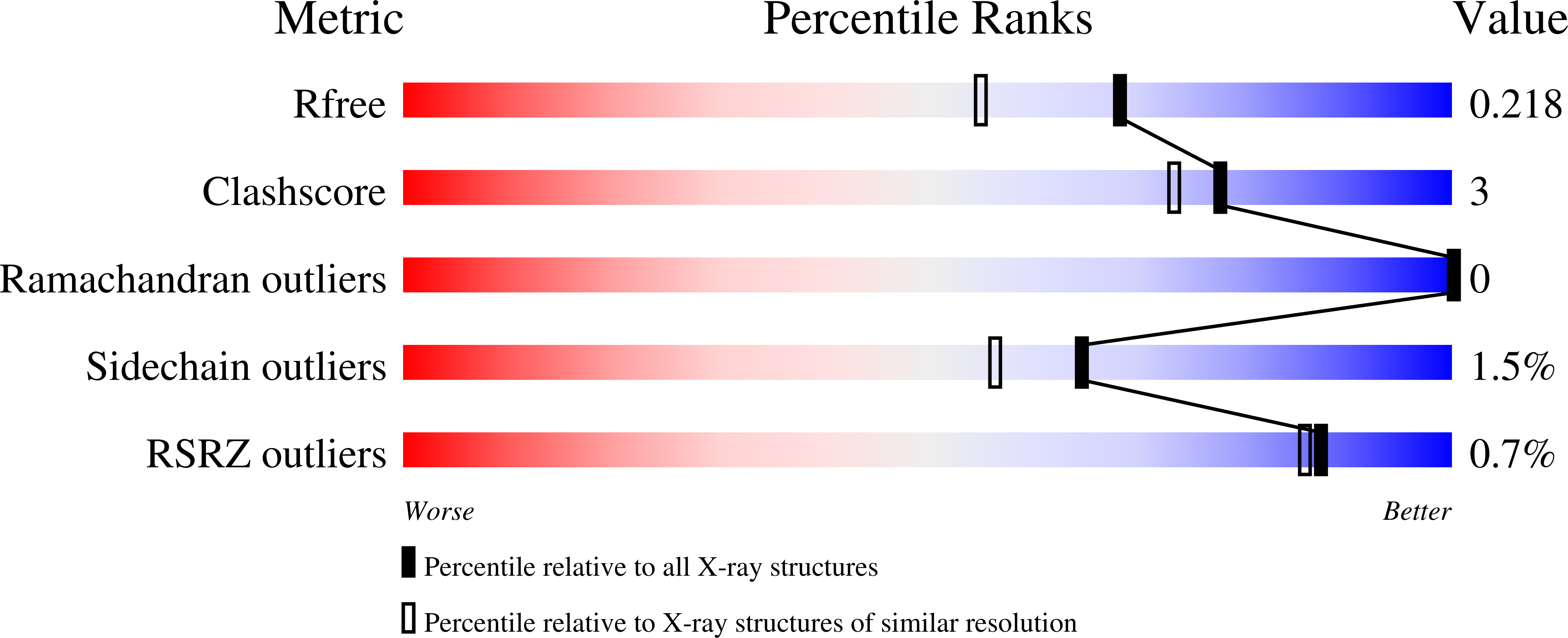
2XTY

Structure of QnrB1 (R167E-Trypsin Treated), a plasmid-mediated fluoroquinolone resistance protein


Experimental Data Snapshot

  • Method: X-RAY DIFFRACTION
  • Resolution: 1.80 Å
  • R-Value Free: 0.221 
  • R-Value Work: 0.180 
  • R-Value Observed: 0.182 

wwPDB Validation   3D Report Full Report


This is version 1.3 of the entry. See complete history


Literature

Structure of Qnrb1, a Plasmid-Mediated Fluoroquinolone Resistance Factor.

Vetting, M.W.Hegde, S.S.Wang, M.Jacoby, G.A.Hooper, D.C.Blanchard, J.S.

(2011) J Biol Chem 286: 25265

  • DOI: https://doi.org/10.1074/jbc.M111.226936
  • Primary Citation of Related Structures:  
    2XTW, 2XTX, 2XTY

  • PubMed Abstract: 

    QnrB1 is a plasmid-encoded pentapeptide repeat protein (PRP) that confers a moderate degree of resistance to fluoroquinolones. Its gene was cloned into an expression vector with an N-terminal polyhistidine tag, and the protein was purified by nickel affinity chromatography. The structure of QnrB1 was determined by a combination of trypsinolysis, surface mutagenesis, and single anomalous dispersion phasing. QnrB1 folds as a right-handed quadrilateral β-helix with a highly asymmetric dimeric structure typical of PRP-topoisomerase poison resistance factors. The threading of pentapeptides into the β-helical fold is interrupted by two noncanonical PRP sequences that produce outward projecting loops that interrupt the regularity of the PRP surface. Deletion of the larger upper loop eliminated the protective effect of QnrB1 on DNA gyrase toward inhibition by quinolones, whereas deletion of the smaller lower loop drastically reduced the protective effect. These loops are conserved among all plasmid-based Qnr variants (QnrA, QnrC, QnrD, and QnrS) and some chromosomally encoded Qnr varieties. A mechanism in which PRP-topoisomerase poison resistance factors bind to and disrupt the quinolone-DNA-gyrase interaction is proposed.


  • Organizational Affiliation

    Department of Biochemistry, Albert Einstein College of Medicine, Bronx, New York 10461, USA.


Macromolecules
Find similar proteins by:  (by identity cutoff)  |  3D Structure
Entity ID: 1
MoleculeChains Sequence LengthOrganismDetailsImage
QNRB1
A, B
217Klebsiella pneumoniaeMutation(s): 1 
UniProt
Find proteins for Q2I1Y8 (Klebsiella pneumoniae)
Explore Q2I1Y8 
Go to UniProtKB:  Q2I1Y8
Entity Groups  
Sequence Clusters30% Identity50% Identity70% Identity90% Identity95% Identity100% Identity
UniProt GroupQ2I1Y8
Sequence Annotations
Expand
  • Reference Sequence
Experimental Data & Validation

Experimental Data

  • Method: X-RAY DIFFRACTION
  • Resolution: 1.80 Å
  • R-Value Free: 0.221 
  • R-Value Work: 0.180 
  • R-Value Observed: 0.182 
  • Space Group: P 41 21 2
Unit Cell:
Length ( Å )Angle ( ˚ )
a = 55.33α = 90
b = 55.33β = 90
c = 282.46γ = 90
Software Package:
Software NamePurpose
PHENIXrefinement
MOSFLMdata reduction
SCALAdata scaling
PHENIXphasing

Structure Validation

View Full Validation Report


Entry History 

Deposition Data

Revision History  (Full details and data files)

  • Version 1.0: 2010-10-27
    Type: Initial release
  • Version 1.1: 2011-07-27
    Changes: Database references, Version format compliance
  • Version 1.2: 2019-05-08
    Changes: Data collection, Experimental preparation, Other
  • Version 1.3: 2023-12-20
    Changes: Data collection, Database references, Other, Refinement description