2X8B

Crystal structure of human acetylcholinesterase inhibited by aged tabun and complexed with fasciculin-II


Experimental Data Snapshot

  • Method: X-RAY DIFFRACTION
  • Resolution: 2.95 Å
  • R-Value Free: 0.253 
  • R-Value Work: 0.183 
  • R-Value Observed: 0.187 

wwPDB Validation   3D Report Full Report


Ligand Structure Quality Assessment 


This is version 1.6 of the entry. See complete history


Literature

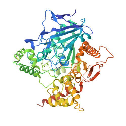
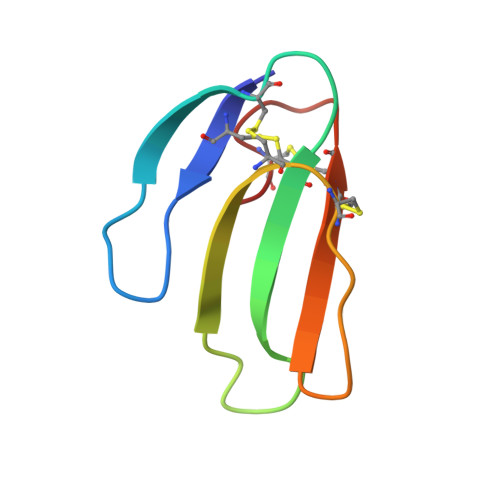
Structural evidence that human acetylcholinesterase inhibited by tabun ages through O-dealkylation.

Carletti, E.Colletier, J.P.Dupeux, F.Trovaslet, M.Masson, P.Nachon, F.

(2010) J Med Chem 53: 4002-4008

  • DOI: https://doi.org/10.1021/jm901853b
  • Primary Citation of Related Structures:  
    2X8B

  • PubMed Abstract: 

    Tabun is a warfare agent that inhibits human acetylcholinesterase (hAChE) by rapid phosphylation of the catalytic serine. A time-dependent reaction occurs on the tabun adduct, leading to an "aged" enzyme, resistant to oxime reactivators. The aging reaction may proceed via either dealkylation or deamidation, depending on the stereochemistry of the phosphoramidyl adduct. We solved the X-ray structure of aged tabun-hAChE complexed with fasciculin II, and we show that aging proceeds through O-dealkylation, in agreement with the aging mechanism that we determined for tabun-inhibited human butyrylcholinesterase and mouse acetylcholinesterase. Noteworthy, aging and binding of fasciculin II lead to an improved thermostability, resulting from additional stabilizing interactions between the two subdomains that face each other across the active site gorge. This first structure of hAChE inhibited by a nerve agent provides structural insight into the inhibition and aging mechanisms and a structural template for the design of molecules capable of reactivating aged hAChE.


  • Organizational Affiliation

    Laboratoire de Biophysique Moléculaire, Institut de Biologie Structurale (CEA-CNRS-UJF), Grenoble, France.


Macromolecules
Find similar proteins by:  (by identity cutoff)  |  3D Structure
Entity ID: 1
MoleculeChains Sequence LengthOrganismDetailsImage
ACETHYLCHOLINESTERASE583Homo sapiensMutation(s): 0 
EC: 3.1.1.7
UniProt & NIH Common Fund Data Resources
Find proteins for P22303 (Homo sapiens)
Explore P22303 
Go to UniProtKB:  P22303
PHAROS:  P22303
GTEx:  ENSG00000087085 
Entity Groups  
Sequence Clusters30% Identity50% Identity70% Identity90% Identity95% Identity100% Identity
UniProt GroupP22303
Sequence Annotations
Expand
  • Reference Sequence
Find similar proteins by:  (by identity cutoff)  |  3D Structure
Entity ID: 2
MoleculeChains Sequence LengthOrganismDetailsImage
FASCICULIN-261Dendroaspis angusticepsMutation(s): 0 
UniProt
Find proteins for P0C1Z0 (Dendroaspis angusticeps)
Explore P0C1Z0 
Go to UniProtKB:  P0C1Z0
Entity Groups  
Sequence Clusters30% Identity50% Identity70% Identity90% Identity95% Identity100% Identity
UniProt GroupP0C1Z0
Sequence Annotations
Expand
  • Reference Sequence
Small Molecules
Ligands 4 Unique
IDChains Name / Formula / InChI Key2D Diagram3D Interactions
NAG
Query on NAG

Download Ideal Coordinates CCD File 
L [auth A],
M [auth A]
2-acetamido-2-deoxy-beta-D-glucopyranose
C8 H15 N O6
OVRNDRQMDRJTHS-FMDGEEDCSA-N
SO4
Query on SO4

Download Ideal Coordinates CCD File 
I [auth A]SULFATE ION
O4 S
QAOWNCQODCNURD-UHFFFAOYSA-L
CL
Query on CL

Download Ideal Coordinates CCD File 
C [auth A]
D [auth A]
E [auth A]
F [auth A]
G [auth A]
C [auth A],
D [auth A],
E [auth A],
F [auth A],
G [auth A],
H [auth A],
N [auth A],
O [auth A],
P [auth A],
Q [auth A],
R [auth A]
CHLORIDE ION
Cl
VEXZGXHMUGYJMC-UHFFFAOYSA-M
UNX
Query on UNX

Download Ideal Coordinates CCD File 
J [auth A],
K [auth A],
S [auth A]
UNKNOWN ATOM OR ION
X
Modified Residues  1 Unique
IDChains TypeFormula2D DiagramParent
SEN
Query on SEN
A
L-PEPTIDE LINKINGC5 H13 N2 O5 PSER
Experimental Data & Validation

Experimental Data

  • Method: X-RAY DIFFRACTION
  • Resolution: 2.95 Å
  • R-Value Free: 0.253 
  • R-Value Work: 0.183 
  • R-Value Observed: 0.187 
  • Space Group: H 3 2
Unit Cell:
Length ( Å )Angle ( ˚ )
a = 151.31α = 90
b = 151.31β = 90
c = 247.24γ = 120
Software Package:
Software NamePurpose
REFMACrefinement
XDSdata reduction
XSCALEdata scaling
XDSphasing

Structure Validation

View Full Validation Report


Ligand Structure Quality Assessment 


Entry History 

Deposition Data

Revision History  (Full details and data files)

  • Version 1.0: 2010-04-28
    Type: Initial release
  • Version 1.1: 2011-06-30
    Changes: Version format compliance
  • Version 1.2: 2011-07-13
    Changes: Version format compliance
  • Version 1.3: 2018-02-28
    Changes: Database references, Source and taxonomy
  • Version 1.4: 2020-07-29
    Type: Remediation
    Reason: Carbohydrate remediation
    Changes: Data collection, Derived calculations, Other, Structure summary
  • Version 1.5: 2021-05-12
    Changes: Derived calculations, Structure summary
  • Version 1.6: 2023-12-20
    Changes: Data collection, Database references, Refinement description