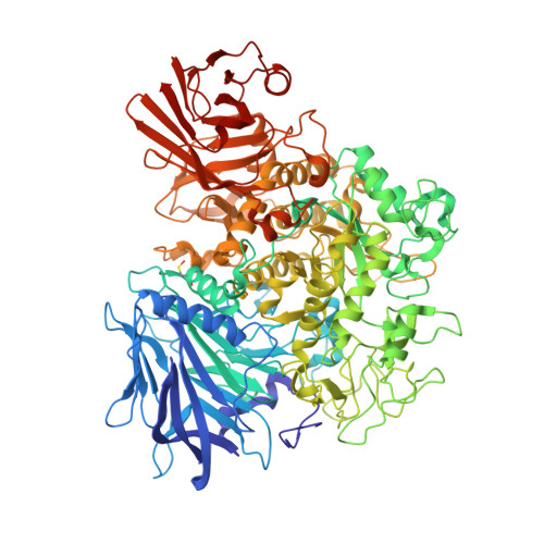
2X2H

Crystal structure of the Gracilariopsis lemaneiformis alpha-1,4- glucan lyase


Experimental Data Snapshot

  • Method: X-RAY DIFFRACTION
  • Resolution: 2.06 Å
  • R-Value Free: 0.215 
  • R-Value Work: 0.168 
  • R-Value Observed: 0.171 

wwPDB Validation   3D Report Full Report


This is version 1.7 of the entry. See complete history


Literature

Crystal Structure of Alpha-1,4-Glucan Lyase, a Unique Glycoside Hydrolase Family Member with a Novel Catalytic Mechanism.

Rozeboom, H.J.Yu, S.Madrid, S.Kalk, K.H.Zhang, R.Dijkstra, B.W.

(2013) J Biol Chem 288: 26764

  • DOI: https://doi.org/10.1074/jbc.M113.485896
  • Primary Citation of Related Structures:  
    2X2H, 2X2J, 4AMW, 4AMX

  • PubMed Abstract: 

    α-1,4-Glucan lyase (EC 4.2.2.13) from the red seaweed Gracilariopsis lemaneiformis cleaves α-1,4-glucosidic linkages in glycogen, starch, and malto-oligosaccharides, yielding the keto-monosaccharide 1,5-anhydro-D-fructose. The enzyme belongs to glycoside hydrolase family 31 (GH31) but degrades starch via an elimination reaction instead of hydrolysis. The crystal structure shows that the enzyme, like GH31 hydrolases, contains a (β/α)8-barrel catalytic domain with B and B' subdomains, an N-terminal domain N, and the C-terminal domains C and D. The N-terminal domain N of the lyase was found to bind a trisaccharide. Complexes of the enzyme with acarbose and 1-dexoynojirimycin and two different covalent glycosyl-enzyme intermediates obtained with fluorinated sugar analogues show that, like GH31 hydrolases, the aspartic acid residues Asp(553) and Asp(665) are the catalytic nucleophile and acid, respectively. However, as a unique feature, the catalytic nucleophile is in a position to act also as a base that abstracts a proton from the C2 carbon atom of the covalently bound subsite -1 glucosyl residue, thus explaining the unique lyase activity of the enzyme. One Glu to Val mutation in the active site of the homologous α-glucosidase from Sulfolobus solfataricus resulted in a shift from hydrolytic to lyase activity, demonstrating that a subtle amino acid difference can promote lyase activity in a GH31 hydrolase.


  • Organizational Affiliation

    From the Laboratory of Biophysical Chemistry, Groningen Biomolecular Sciences and Biotechnology Institute, University of Groningen, Nijenborgh 7, 9747 AG Groningen, The Netherlands.


Macromolecules
Find similar proteins by:  (by identity cutoff)  |  3D Structure
Entity ID: 1
MoleculeChains Sequence LengthOrganismDetailsImage
ALPHA-1,4-GLUCAN LYASE ISOZYME 1
A, B, C, D
1,027Gracilariopsis lemaneiformisMutation(s): 0 
EC: 4.2.2.13
UniProt
Find proteins for Q9STC1 (Gracilariopsis lemaneiformis)
Explore Q9STC1 
Go to UniProtKB:  Q9STC1
Entity Groups  
Sequence Clusters30% Identity50% Identity70% Identity90% Identity95% Identity100% Identity
UniProt GroupQ9STC1
Sequence Annotations
Expand
  • Reference Sequence
Small Molecules
Ligands 3 Unique
IDChains Name / Formula / InChI Key2D Diagram3D Interactions
GOL
Query on GOL

Download Ideal Coordinates CCD File 
AA [auth D]
BA [auth D]
CA [auth D]
E [auth A]
F [auth A]
AA [auth D],
BA [auth D],
CA [auth D],
E [auth A],
F [auth A],
G [auth A],
H [auth A],
I [auth A],
J [auth A],
K [auth A],
Q [auth B],
R [auth B],
S [auth B],
U [auth C],
V [auth C],
W [auth C]
GLYCEROL
C3 H8 O3
PEDCQBHIVMGVHV-UHFFFAOYSA-N
ACT
Query on ACT

Download Ideal Coordinates CCD File 
DA [auth D]
EA [auth D]
L [auth A]
M [auth A]
N [auth A]
DA [auth D],
EA [auth D],
L [auth A],
M [auth A],
N [auth A],
O [auth A],
X [auth C],
Y [auth C],
Z [auth C]
ACETATE ION
C2 H3 O2
QTBSBXVTEAMEQO-UHFFFAOYSA-M
CL
Query on CL

Download Ideal Coordinates CCD File 
P [auth A],
T [auth B]
CHLORIDE ION
Cl
VEXZGXHMUGYJMC-UHFFFAOYSA-M
Modified Residues  1 Unique
IDChains TypeFormula2D DiagramParent
CSO
Query on CSO
A, B, C, D
L-PEPTIDE LINKINGC3 H7 N O3 SCYS
Experimental Data & Validation

Experimental Data

  • Method: X-RAY DIFFRACTION
  • Resolution: 2.06 Å
  • R-Value Free: 0.215 
  • R-Value Work: 0.168 
  • R-Value Observed: 0.171 
  • Space Group: P 1
Unit Cell:
Length ( Å )Angle ( ˚ )
a = 91.678α = 80.54
b = 96.629β = 83.28
c = 134.457γ = 85.26
Software Package:
Software NamePurpose
REFMACrefinement
DENZOdata reduction
SCALEPACKdata scaling
PHASERphasing

Structure Validation

View Full Validation Report


Entry History 

Deposition Data

Revision History  (Full details and data files)

  • Version 1.0: 2011-01-19
    Type: Initial release
  • Version 1.1: 2013-08-14
    Changes: Database references, Non-polymer description, Refinement description, Version format compliance
  • Version 1.2: 2013-09-25
    Changes: Database references
  • Version 1.3: 2019-01-30
    Changes: Data collection, Experimental preparation, Other
  • Version 1.4: 2019-02-06
    Changes: Data collection, Experimental preparation
  • Version 1.5: 2019-07-10
    Changes: Data collection, Derived calculations
  • Version 1.6: 2019-07-24
    Changes: Data collection
  • Version 1.7: 2023-12-20
    Changes: Data collection, Database references, Derived calculations, Other, Refinement description