2WY7

Staphylococcus aureus complement subversion protein Sbi-IV in complex with complement fragment C3d revealing an alternative binding mode


Experimental Data Snapshot

  • Method: X-RAY DIFFRACTION
  • Resolution: 1.70 Å
  • R-Value Free: 0.205 
  • R-Value Work: 0.172 
  • R-Value Observed: 0.174 

wwPDB Validation   3D Report Full Report


This is version 1.2 of the entry. See complete history


Literature

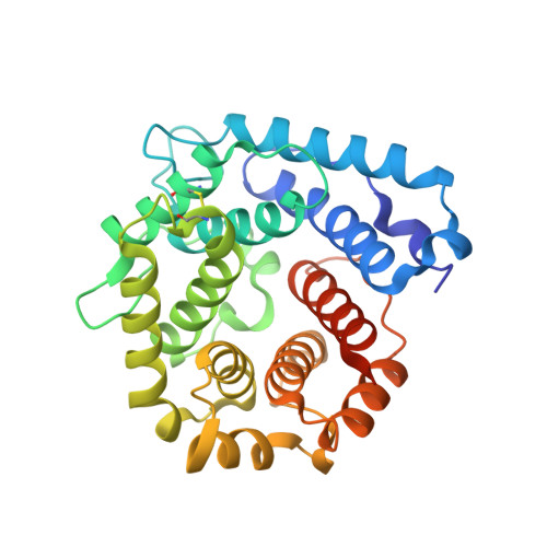
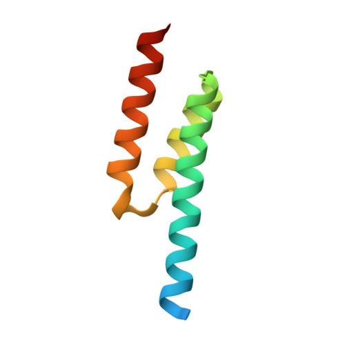
A Structural Basis for Staphylococcal Complement Subversion: X-Ray Structure of the Complement- Binding Domain of Staphylococcus Aureus Protein Sbi in Complex with Ligand C3D.

Clark, E.A.Crennell, S.Upadhyay, A.Zozulya, A.V.Mackay, J.D.Svergun, D.I.Bagby, S.Van Den Elsen, J.M.

(2011) Mol Immunol 48: 452

  • DOI: https://doi.org/10.1016/j.molimm.2010.09.017
  • Primary Citation of Related Structures:  
    2WY7, 2WY8

  • PubMed Abstract: 

    The structure of the complement-binding domain of Staphylococcus aureus protein Sbi (Sbi-IV) in complex with ligand C3d is presented. The 1.7Å resolution structure reveals the molecular details of the recognition of thioester-containing fragment C3d of the central complement component C3, involving interactions between residues of Sbi-IV helix α2 and the acidic concave surface of C3d. The complex provides a structural basis for the binding preference of Sbi for native C3 over C3b and explains how Sbi-IV inhibits the interaction between C3d and complement receptor 2. A second C3d binding site on Sbi-IV is identified in the crystal structure that is not observed in related S. aureus C3 inhibitors Efb-C and Ehp. This binding mode perhaps hints as to how Sbi-IV, as part of Sbi, forms a C3b-Sbi adduct and causes futile consumption of C3, an extraordinary aspect of Sbi function that is not shared by any other known Staphylococcal complement inhibitor.


  • Organizational Affiliation

    University of Bath, Department of Biology and Biochemistry, Claverton Down, Bath BA2 7AY, United Kingdom.


Macromolecules
Find similar proteins by:  (by identity cutoff)  |  3D Structure
Entity ID: 1
MoleculeChains Sequence LengthOrganismDetailsImage
COMPLEMENT C3D FRAGMENT310Homo sapiensMutation(s): 1 
UniProt & NIH Common Fund Data Resources
Find proteins for P01024 (Homo sapiens)
Explore P01024 
Go to UniProtKB:  P01024
PHAROS:  P01024
GTEx:  ENSG00000125730 
Entity Groups  
Sequence Clusters30% Identity50% Identity70% Identity90% Identity95% Identity100% Identity
UniProt GroupP01024
Sequence Annotations
Expand
  • Reference Sequence
Find similar proteins by:  (by identity cutoff)  |  3D Structure
Entity ID: 2
MoleculeChains Sequence LengthOrganismDetailsImage
IGG-BINDING PROTEINB [auth Q]80Staphylococcus aureusMutation(s): 0 
Entity Groups  
Sequence Clusters30% Identity50% Identity70% Identity90% Identity95% Identity100% Identity
Sequence Annotations
Expand
  • Reference Sequence
Experimental Data & Validation

Experimental Data

  • Method: X-RAY DIFFRACTION
  • Resolution: 1.70 Å
  • R-Value Free: 0.205 
  • R-Value Work: 0.172 
  • R-Value Observed: 0.174 
  • Space Group: P 21 21 21
Unit Cell:
Length ( Å )Angle ( ˚ )
a = 53.43α = 90
b = 81.01β = 90
c = 87.83γ = 90
Software Package:
Software NamePurpose
REFMACrefinement
HKL-2000data reduction
DENZOdata scaling
BALBESphasing

Structure Validation

View Full Validation Report


Entry History 

Deposition Data

Revision History  (Full details and data files)

  • Version 1.0: 2010-12-01
    Type: Initial release
  • Version 1.1: 2013-09-18
    Changes: Derived calculations, Non-polymer description, Version format compliance
  • Version 1.2: 2023-12-20
    Changes: Data collection, Database references, Derived calculations, Other, Refinement description