2W54

Crystal Structure of Xanthine Dehydrogenase from Rhodobacter capsulatus in Complex with Bound Inhibitor Pterin-6-aldehyde


Experimental Data Snapshot

  • Method: X-RAY DIFFRACTION
  • Resolution: 3.30 Å
  • R-Value Free: 0.224 
  • R-Value Work: 0.186 
  • R-Value Observed: 0.188 

wwPDB Validation   3D Report Full Report


Ligand Structure Quality Assessment 


This is version 1.4 of the entry. See complete history


Literature

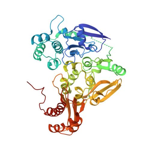
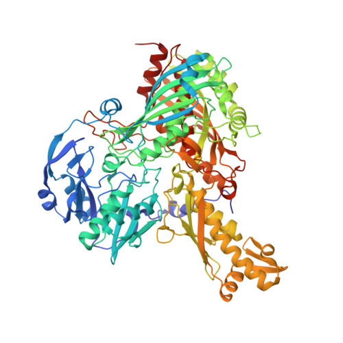
Mechanism of Substrate and Inhibitor Binding of Rhodobacter Capsulatus Xanthine Dehydrogenase.

Dietzel, U.Kuper, J.Doebbler, J.A.Schulte, A.Truglio, J.J.Leimkuhler, S.Kisker, C.

(2009) J Biol Chem 284: 8768

  • DOI: https://doi.org/10.1074/jbc.M808114200
  • Primary Citation of Related Structures:  
    2W3R, 2W3S, 2W54, 2W55

  • PubMed Abstract: 

    Rhodobacter capsulatus xanthine dehydrogenase (XDH) is an (alphabeta)(2) heterotetrameric cytoplasmic enzyme that resembles eukaryotic xanthine oxidoreductases in respect to both amino acid sequence and structural fold. To obtain a detailed understanding of the mechanism of substrate and inhibitor binding at the active site, we solved crystal structures of R. capsulatus XDH in the presence of its substrates hypoxanthine, xanthine, and the inhibitor pterin-6-aldehyde using either the inactive desulfo form of the enzyme or an active site mutant (E(B)232Q) to prevent substrate turnover. The hypoxanthine- and xanthine-bound structures reveal the orientation of both substrates at the active site and show the importance of residue Glu(B)-232 for substrate positioning. The oxygen atom at the C-6 position of both substrates is oriented toward Arg(B)-310 in the active site. Thus the substrates bind in an orientation opposite to the one seen in the structure of the reduced enzyme with the inhibitor oxypurinol. The tightness of the substrates in the active site suggests that the intermediate products must exit the binding pocket to allow first the attack of the C-2, followed by oxidation of the C-8 atom to form the final product uric acid. Structural studies of pterin-6-aldehyde, a potent inhibitor of R. capsulatus XDH, contribute further to the understanding of the relative positioning of inhibitors and substrates in the binding pocket. Steady state kinetics reveal a competitive inhibition pattern with a K(i) of 103.57 +/- 18.96 nm for pterin-6-aldehyde.


  • Organizational Affiliation

    Rudolf Virchow Center for Experimental Biomedicine, Institute for Structural Biology, University of Würzburg, Versbacher Strasse 9, 97078 Würzburg, Germany.


Macromolecules
Find similar proteins by:  (by identity cutoff)  |  3D Structure
Entity ID: 1
MoleculeChains Sequence LengthOrganismDetailsImage
XANTHINE DEHYDROGENASE
A, C, E, G
462Rhodobacter capsulatusMutation(s): 0 
EC: 1.1.1.204
UniProt
Find proteins for O54050 (Rhodobacter capsulatus)
Explore O54050 
Go to UniProtKB:  O54050
Entity Groups  
Sequence Clusters30% Identity50% Identity70% Identity90% Identity95% Identity100% Identity
UniProt GroupO54050
Sequence Annotations
Expand
  • Reference Sequence
Find similar proteins by:  (by identity cutoff)  |  3D Structure
Entity ID: 2
MoleculeChains Sequence LengthOrganismDetailsImage
XANTHINE DEHYDROGENASE
B, D, F, H
777Rhodobacter capsulatusMutation(s): 0 
EC: 1.1.1.204
UniProt
Find proteins for O54051 (Rhodobacter capsulatus)
Explore O54051 
Go to UniProtKB:  O54051
Entity Groups  
Sequence Clusters30% Identity50% Identity70% Identity90% Identity95% Identity100% Identity
UniProt GroupO54051
Sequence Annotations
Expand
  • Reference Sequence
Small Molecules
Ligands 5 Unique
IDChains Name / Formula / InChI Key2D Diagram3D Interactions
FAD
Query on FAD

Download Ideal Coordinates CCD File 
CA [auth G],
K [auth A],
Q [auth C],
W [auth E]
FLAVIN-ADENINE DINUCLEOTIDE
C27 H33 N9 O15 P2
VWWQXMAJTJZDQX-UYBVJOGSSA-N
XAX
Query on XAX

Download Ideal Coordinates CCD File 
DA [auth H],
L [auth B],
R [auth D],
X [auth F]
{[(5aR,8R,9aR)-2-amino-4-oxo-6,7-di(sulfanyl-kappaS)-3,5,5a,8,9a,10-hexahydro-4H-pyrano[3,2-g]pteridin-8-yl]methyl dihydrogenato(2-) phosphate}(hydroxy)oxo(thioxo)molybdenum
C10 H13 Mo N5 O8 P S3
OIQYCPXIZLGKQT-BKZHXLINSA-K
HHR
Query on HHR

Download Ideal Coordinates CCD File 
FA [auth H],
N [auth B],
T [auth D],
Z [auth F]
6-HYDROXYMETHYLPTERIN
C7 H7 N5 O2
XGWIBNWDLMIPNF-UHFFFAOYSA-N
FES
Query on FES

Download Ideal Coordinates CCD File 
AA [auth G]
BA [auth G]
I [auth A]
J [auth A]
O [auth C]
AA [auth G],
BA [auth G],
I [auth A],
J [auth A],
O [auth C],
P [auth C],
U [auth E],
V [auth E]
FE2/S2 (INORGANIC) CLUSTER
Fe2 S2
NIXDOXVAJZFRNF-UHFFFAOYSA-N
BA
Query on BA

Download Ideal Coordinates CCD File 
EA [auth H],
M [auth B],
S [auth D],
Y [auth F]
BARIUM ION
Ba
XDFCIPNJCBUZJN-UHFFFAOYSA-N
Binding Affinity Annotations 
IDSourceBinding Affinity
HHR PDBBind:  2W54 Ki: 103.6 (nM) from 1 assay(s)
Experimental Data & Validation

Experimental Data

  • Method: X-RAY DIFFRACTION
  • Resolution: 3.30 Å
  • R-Value Free: 0.224 
  • R-Value Work: 0.186 
  • R-Value Observed: 0.188 
  • Space Group: P 1
Unit Cell:
Length ( Å )Angle ( ˚ )
a = 92.571α = 109.63
b = 140.695β = 105.83
c = 157.859γ = 101.23
Software Package:
Software NamePurpose
REFMACrefinement
HKL-2000data reduction
HKL-2000data scaling
PHASERphasing

Structure Validation

View Full Validation Report


Ligand Structure Quality Assessment 


Entry History 

Deposition Data

Revision History  (Full details and data files)

  • Version 1.0: 2008-12-23
    Type: Initial release
  • Version 1.1: 2011-07-13
    Changes: Advisory, Version format compliance
  • Version 1.2: 2015-04-22
    Changes: Atomic model, Derived calculations, Non-polymer description, Other, Structure summary
  • Version 1.3: 2019-05-08
    Changes: Data collection, Experimental preparation, Other
  • Version 1.4: 2023-12-13
    Changes: Data collection, Database references, Derived calculations, Other, Refinement description