Solution Structure of the Dimerization Domain of Ribosomal Protein P2 Provides Insights for the Structural Organization of Eukaryotic Stalk.
Lee, K.M., Yu, C.W., Chan, D.S., Chiu, T.Y., Zhu, G., Sze, K.H., Shaw, P.C., Wong, K.B.(2010) Nucleic Acids Res 38: 5206
- PubMed: 20385603
- DOI: https://doi.org/10.1093/nar/gkq231
- Primary Citation of Related Structures:
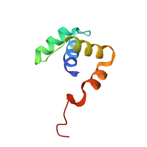
2W1O - PubMed Abstract:
The lateral stalk of ribosome is responsible for kingdom-specific binding of translation factors and activation of GTP hydrolysis that drives protein synthesis. In eukaryotes, the stalk is composed of acidic ribosomal proteins P0, P1 and P2 that constitute a pentameric P-complex in 1: 2: 2 ratio. We have determined the solution structure of the N-terminal dimerization domain of human P2 (NTD-P2), which provides insights into the structural organization of the eukaryotic stalk. Our structure revealed that eukaryotic stalk protein P2 forms a symmetric homodimer in solution, and is structurally distinct from the bacterial counterpart L12 homodimer. The two subunits of NTD-P2 form extensive hydrophobic interactions in the dimeric interface that buries 2400 A(2) of solvent accessible surface area. We have showed that P1 can dissociate P2 homodimer spontaneously to form a more stable P1/P2 1 : 1 heterodimer. By homology modelling, we identified three exposed polar residues on helix-3 of P2 are substituted by conserved hydrophobic residues in P1. Confirmed by mutagenesis, we showed that these residues on helix-3 of P1 are not involved in the dimerization of P1/P2, but instead play a vital role in anchoring P1/P2 heterodimer to P0. Based on our results, models of the eukaryotic stalk complex were proposed.
Organizational Affiliation:
Department of Biochemistry, Centre for Protein Science and Crystallography, The Chinese University of Hong Kong, Hong Kong, China.