2VQB

Bacterial flavin-containing monooxygenase in complex with NADP: soaking in aerated solution


Experimental Data Snapshot

  • Method: X-RAY DIFFRACTION
  • Resolution: 2.80 Å
  • R-Value Free: 0.242 
  • R-Value Work: 0.226 
  • R-Value Observed: 0.226 

wwPDB Validation   3D Report Full Report


Ligand Structure Quality Assessment 


This is version 1.3 of the entry. See complete history


Literature

Revealing the Moonlighting Role of Nadp in the Structure of a Flavin-Containing Monooxygenase.

Alfieri, A.Malito, E.Orru, R.Fraaije, M.W.Mattevi, A.

(2008) Proc Natl Acad Sci U S A 105: 6572

  • DOI: https://doi.org/10.1073/pnas.0800859105
  • Primary Citation of Related Structures:  
    2VQ7, 2VQB

  • PubMed Abstract: 

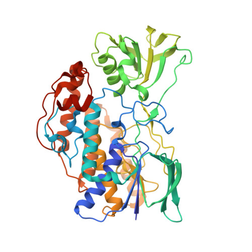
    Flavin-containing monooxygenases (FMOs) are, after cytochromes P450, the most important monooxygenase system in humans and are involved in xenobiotics metabolism and variability in drug response. The x-ray structure of a soluble prokaryotic FMO from Methylophaga sp. strain SK1 has been solved at 2.6-A resolution and is now the protein of known structure with the highest sequence similarity to human FMOs. The structure possesses a two-domain architecture, with both FAD and NADP(+) well defined by the electron density maps. Biochemical analysis shows that the prokaryotic enzyme shares many functional properties with mammalian FMOs, including substrate specificity and the ability to stabilize the hydroperoxyflavin intermediate that is crucial in substrate oxygenation. On the basis of their location in the structure, the nicotinamide ring and the adjacent ribose of NADP(+) turn out to be an integral part of the catalytic site being actively engaged in the stabilization of the oxygenating intermediate. This feature suggests that NADP(H) has a moonlighting role, in that it adopts two binding modes that allow it to function in both flavin reduction and oxygen reactivity modulation, respectively. We hypothesize that a relative domain rotation is needed to bring NADP(H) to these distinct positions inside the active site. Localization of mutations in human FMO3 that are known to cause trimethylaminuria (fish-odor syndrome) in the elucidated FMO structure provides a structural explanation for their biological effects.


  • Organizational Affiliation

    Department of Genetics and Microbiology, University of Pavia, Via Ferrata 1, 27100 Pavia, Italy.


Macromolecules
Find similar proteins by:  (by identity cutoff)  |  3D Structure
Entity ID: 1
MoleculeChains Sequence LengthOrganismDetailsImage
FLAVIN-CONTAINING MONOOXYGENASE
A, B, C, D
461Methylophaga aminisulfidivoransMutation(s): 2 
UniProt
Find proteins for Q83XK4 (Methylophaga aminisulfidivorans)
Explore Q83XK4 
Go to UniProtKB:  Q83XK4
Entity Groups  
Sequence Clusters30% Identity50% Identity70% Identity90% Identity95% Identity100% Identity
UniProt GroupQ83XK4
Sequence Annotations
Expand
  • Reference Sequence
Small Molecules
Ligands 5 Unique
IDChains Name / Formula / InChI Key2D Diagram3D Interactions
FAD
Query on FAD

Download Ideal Coordinates CCD File 
E [auth A],
K [auth B],
Q [auth C],
W [auth D]
FLAVIN-ADENINE DINUCLEOTIDE
C27 H33 N9 O15 P2
VWWQXMAJTJZDQX-UYBVJOGSSA-N
NAP
Query on NAP

Download Ideal Coordinates CCD File 
F [auth A],
L [auth B],
R [auth C],
X [auth D]
NADP NICOTINAMIDE-ADENINE-DINUCLEOTIDE PHOSPHATE
C21 H28 N7 O17 P3
XJLXINKUBYWONI-NNYOXOHSSA-N
EPE
Query on EPE

Download Ideal Coordinates CCD File 
H [auth A],
N [auth B],
S [auth C],
Z [auth D]
4-(2-HYDROXYETHYL)-1-PIPERAZINE ETHANESULFONIC ACID
C8 H18 N2 O4 S
JKMHFZQWWAIEOD-UHFFFAOYSA-N
CL
Query on CL

Download Ideal Coordinates CCD File 
G [auth A],
M [auth B],
V [auth C],
Y [auth D]
CHLORIDE ION
Cl
VEXZGXHMUGYJMC-UHFFFAOYSA-M
OXY
Query on OXY

Download Ideal Coordinates CCD File 
AA [auth D]
BA [auth D]
I [auth A]
J [auth A]
O [auth B]
AA [auth D],
BA [auth D],
I [auth A],
J [auth A],
O [auth B],
P [auth B],
T [auth C],
U [auth C]
OXYGEN MOLECULE
O2
MYMOFIZGZYHOMD-UHFFFAOYSA-N
Experimental Data & Validation

Experimental Data

  • Method: X-RAY DIFFRACTION
  • Resolution: 2.80 Å
  • R-Value Free: 0.242 
  • R-Value Work: 0.226 
  • R-Value Observed: 0.226 
  • Space Group: P 61
Unit Cell:
Length ( Å )Angle ( ˚ )
a = 218.665α = 90
b = 218.665β = 90
c = 130.676γ = 120
Software Package:
Software NamePurpose
REFMACrefinement
MOSFLMdata reduction
SCALAdata scaling
CCP4phasing

Structure Validation

View Full Validation Report


Ligand Structure Quality Assessment 


Entry History 

Deposition Data

Revision History  (Full details and data files)

  • Version 1.0: 2008-04-22
    Type: Initial release
  • Version 1.1: 2011-05-08
    Changes: Version format compliance
  • Version 1.2: 2011-07-13
    Changes: Version format compliance
  • Version 1.3: 2023-12-13
    Changes: Data collection, Database references, Derived calculations, Other, Refinement description